موقع د. كمال سيد الدراوي
طبي_ اكاديمي _ ثقافي _ تعليمي _ _ استشارات طبية_فيديو طبي
|
| | | Abdominal Ultrasound |  |
| | |
| كاتب الموضوع | رسالة |
|---|
د.كمال سيد
Admin


عدد المساهمات : 2679
نقاط : 4483
السٌّمعَة : 9
الجنس : 
علم بلدك : 
تاريخ الميلاد : 03/04/1950
تاريخ التسجيل : 30/07/2012
العمر : 74
الموقع : السودان - سنار
العمل/الترفيه : طبيب عمومى وموجات صوتية
الساعة الان :
دعائي : 
|  موضوع: spleen contd موضوع: spleen contd  السبت أكتوبر 05, 2024 6:47 pm السبت أكتوبر 05, 2024 6:47 pm | |
| spleen contdSplenomegalyEnlargement of the spleen is a highly non-specific sign associated with numerous conditions, the most common being 1.infection, 2.portal hypertension, 3.hematological disorders and 4.neoplastic conditions (Box 6.1) Examples of causes of splenomegaly* Portal hypertension* Acute or chronic systemic infection – e.g. hepatitis, AIDS, infectious mononucleosis, sepsis* Haemolytic anaemia, sickle cell disease, thalassaemia, pernicious anaemia, spherocytosis* Malignancy – leukaemia, Hodgkin’s and non-Hodgkin’s lymphoma, myeloproliferative disorders* Storage disorders* Immunological diseasesAs with the liver, measurement of splenic volume is unreliable and not reproducible, due to+ variation in *shape and *subdiaphragmatic access. However, *the length of the spleen is an adequateindicator of size for most purposes and provides a useful baseline for monitoring changes in disease status. The length *(infero-superior) of the normal adult spleen is less than 12 cm.The spleen enlarges *downwards and *medially. Its inferior margin *becomes rounded (Figs 6.1D, 6.2A) and it may *extend below the left kidney and *into the pelvis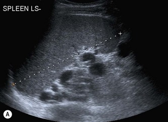 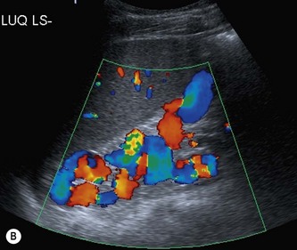 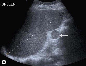 (A) Splenomegaly in portal hypertension.
The inferior splenic margin is blunted, descending below and medial to the left kidney.
Varices are present around the hilum
B/ Varices, in the form of a spleno-renal shunt, in portal hypertension.
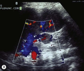
C/ A splenunculus (arrow) at the hilum of a mildly enlarged spleen.
D/ The circulation of the splenunculus derives from the main splenic artery and drains into the main splenic vein.
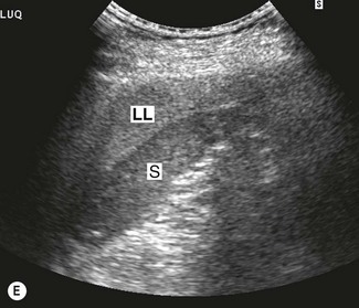
(E) The left lobe of the liver, LL, extends across the abdomen and above the spleen, S, in hepatomegaly,
giving the appearance of a well-defined splenic ‘mass’.
Although the etiology of splenomegaly may not be obvious on ultrasound, the causes can be+
narrowed down by considering the clinical picture and by identifying other relevant appearances
in the abdomen.
Splenomegaly due to portal hypertension, for example, is frequently accompanied by other associated
Splenunculi
In around *10% of the population, a small accessory spleen, or splenunculus, may be located at the *splenic hilum.
These 1.small, 2.well-defined 3.ectopic nodules of 4.splenic tissue (Fig. 6.2C) 5.rarely exceed 2 cm in diameter.
Splenunculi enlarge under the *same circumstances as those which cause splenomegaly, and may also
hypertrophy in post-splenectomy patients*.
The importance of recognizing these lies in differentiating them from 1.lymph nodes, 2.left adrenal nodules
or 3.masses in the tail of pancreas. 4.Colour Doppler may identify the vascular supply as being common
to the main spleen (Fig. 6.2D).
Malignant splenic disease
Lymphoproliferative disorders
These are malignant haematologic conditions, comprising *Hodgkin’s and *non-Hodgkin’s lymphoma
leukaemias and *myeloma*
Lymphoma is the most common malignant disease affecting the spleen (Fig. 6.3). Malignant cells can+
infiltrate the 1.spleen, 2.lymph nodes, 3.bone marrow and *thymus and can also involve the *liver, gastrointestinal tract, *kidney and *other organs. Approximately 3% of malignant diseases are lymphomas*
splenic involvement may be found in up to 60% of lymphomas as a result of dissemination of the disease. *Primary splenic lymphoma, limited to the spleen, is very rare, and accounts for less than 1% of lymphomas
Lymphoma is also *associated with AIDS and infection with HIV has given rise to a broad spectrum of lymphomatous conditions which may be demonstrated on US and CT.
These include masses in the liver, spleen, kidneys, adrenal gland, bowel and other retroperitoneal
and nodal masses.
In addition, the increased use of immunosuppression in transplant patients, and the increased
survival in this group, has also been the cause of an increased incidence of immunodeficiency-related
lymphoma known as PTLD (Fig. 6.3D) (see also Chapter 4)
Post-Transplant Lymphoproliferative Disorders (PTLD) are lymphoid and/OR plasmacytic proliferations
which are types of lymphoma that can happen in pts after having transplant & receiving chronic immunosuppressive therapy
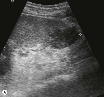
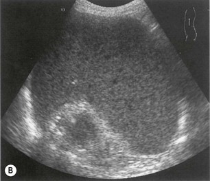
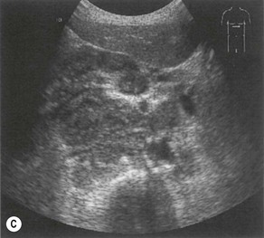
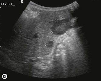
fig. 6.3 • Lymphoma:
(A) A hypoechoic focal lesion in a normal-sized spleen in a patient with AIDS is suspicious for lymphoma.
(B) Enlarged, coarse-textured spleen containing multiple tiny lymphomatous lesions.
(C) Extensive lymphadenopathy in the epigastric region.
(D) hypoechoic focal lesions in a liver transplant
| |
| | | | د.كمال سيد
Admin
عدد المساهمات : 2679
نقاط : 4483
السٌّمعَة : 9
الجنس :
علم بلدك :
تاريخ الميلاد : 03/04/1950
تاريخ التسجيل : 30/07/2012
العمر : 74
الموقع : السودان - سنار
العمل/الترفيه : طبيب عمومى وموجات صوتية
الساعة الان :
دعائي :
| | موضوع: Spleen scanning protocol الجمعة أكتوبر 11, 2024 7:12 pm | |
| Spleen scanning protocol Spleen anatomy Except for its medial hilum, the spleen is +intraperitoneal i.e enclosed in the sac formed by the parietal* peritoneum in the +posterolateral section of the left upper quadrant (left hypochondrium), +beneath the ninth to eleventh intercostal spaces.* The spleen lies posterior and lateral to the +tail of the pancreas, +fundus and body of the stomach and +posterior to the splenic flexure (sharp bend between the transverse and descending colon in the (left upper quadrant, anterior to the spleen .* The medial portion of the spleen is superior and lateral to the left kidney.* The spleen lies +inferior and adjacent to the diaphragm, putting it in close proximity to the +left pleural cavity.* The shape of the spleen is variable, but it is basically ovoid and may resemble a half moon or crescent. Thus, its superoposterior surface (adjacent to the diaphragm) is convex and smooth and its inferomedial surface (in contact with the stomach, left kidney, splenic flexure, pancreas tail) is concave and indented or nodulous. The splenic hilum (entry and exit route for the arterial, venous, and lymphatic vessels and nerves) is located within this concavity. Note that if the spleen becomes enlarged, it loses its concave appearance and becomes more round.* The splenic vein exits the hilum and heads across the body toward the midline, running behind i.e (posterior to) the tail and body of the pancreas, to join the superior mesenteric vein at a point just posterior to the pancreas neck to form the portal vein. This location is commonly referred to as the portal-splenic confluence.* The spleen receives blood from the splenic artery that branches from the celiac trunk of the abdominal aorta and runs left lateral and just superior to the body and tail of the pancreas, to enter the splenic hilum. In many cases, the artery may divide into two or three smaller branches before entering the spleen.
Physiology* Although not essential to life, the spleen 1.filters foreign material from the blood and 2.forms antibodies.* The spleen is a large mass of lymphatic tissue (reticular connective tissue) that is part of the reticuloendothelial system, 3.an essential part of the immune system. It is composed of +phagocytic cells capable of +consuming substances, such as bacteria and viruses making them incapable of causing harm to the body. They also +engulf old cells and abnormal cells, clearing the body of their harmful presence.* The spleen also 4.breaks down hemoglobin, is a blood reservoir, and is important for hematopoiesis i.e (red blood cell formation). in the fetus or when there is severe anemia i.e marked loss of the number of red blood cells produced in bone marrow; symptoms may include fatigue, shortness of breath, irregular heartbeat .Sonographic Appearance* The normal adult spleen appears 1.homogeneous, with 2.medium-level echoes and 3.even texture that is described as isosonic (Isoechoic) or slightly hypoechoic when compared to the normal liver, and hyperechoic relative to kidney parenchyma. In some cases, 4.small vascular branches can be seen interspersed within the spleen. They appear as +anechoic, +round or +tubular structures. Arterial 5.walls usually appear +brighter than venous walls; however, the larger venous structures can clearly be distinguished from the smaller arterial branches at the level of the splenic hilum. Occasionally, 6.small, bright reflections may be visualized throughout the spleen that represent calcified +granulomatous inclusions or +calcifications of small arterial walls.* The 7.orientation of the spleen is +vertically +obliqued in the body, therefore longitudinal and long axis views are seen in +oblique sagittal scanning planes and oblique coronal scanning planes as demonstrated in the image on the following page. This normal spleen appears 8.crescent in shape and + hyperechoic compared to the adjacent left kidney. Notice the spleen’s +smooth outer convexity and its relationship to the diaphragm and pleural space superiorly.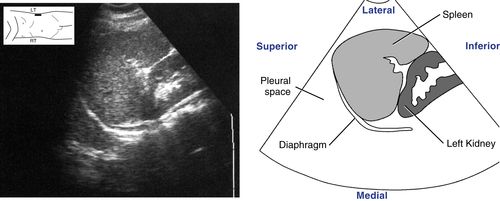
• Axial sections of the spleen are visualized in transverse scanning planes. The following is a transverse scanning plane image from a left lateral approach. It demonstrates an axial section of the spleen that includes the splenic hilum. Note the normal homogeneous appearance and even texture of the spleen. No Preparation needed A 5.0 MHz Transducer for intercostal or lateral subcostal scanning approaches .
A 3 OR 3.5 MHz for anterior or posterior scanning approaches .Breathing Technique Deep, held inspiration. Deep inspiration causes the spleen to descend, making it easier to visualize sonographically. Different breathing techniques should be used whenever the suggested breathing technique does not produce the desired results.
Patient Position Right lateral decubitus. Supine, sitting semierect to erect, and prone as neededSpleen | |
| | | | د.كمال سيد
Admin
عدد المساهمات : 2679
نقاط : 4483
السٌّمعَة : 9
الجنس :
علم بلدك :
تاريخ الميلاد : 03/04/1950
تاريخ التسجيل : 30/07/2012
العمر : 74
الموقع : السودان - سنار
العمل/الترفيه : طبيب عمومى وموجات صوتية
الساعة الان :
دعائي :
| | موضوع: Renal scanning protocol الإثنين أكتوبر 14, 2024 8:32 pm | |
| Renal scanning protocol
* The right kidney is normally situated lower than the left kidney.* The right kidney is posteroinferior to the liver and gallbladder.* The left kidney is inferior and medial to the spleen.* Located immediately superior, anterior, and medial to each kidney is an adrenal gland.Anatomy* The kidneys are described as having a superior and inferior pole, medial and lateral margins and an anterior and posterior surface.* The kidneys are encapsulated by a fibrous renal or true capsule. * Fat surrounds the encapsulated kidneys in the perinephric or perirenal space which is part of the retroperitoneal space within the renal fascia that contains the 1.kidney, 2.perirenal fat and 3.adrenal gland, & 4.proximal ureter * Gerota’s fascia is a fibrous sheath that encloses each 1.kidney, 2.perinephric fat, 3.and adrenal gland.* Normal adult kidneys measure 9 to 12|cm long, 2.5 to 3.5|cm thick, and 4 to 5|cm wide.* Each kidney is composed of two distinct areas, the renal sinus and renal parenchyma:1/ Renal sinus: is the internal cavity within the kidney, which contains the upper expanded part of the ureter called the renal pelvis consists of the +renal hilum and +collecting system A. Renal hilum:1. Hilum is the Medial opening into the renal sinus at the mid portion of the kidney.2. The Hilum transmits, from anterior to posterior, Renal VEIN, Renal Artery, & URETER (VAU). Hilum also transmits sympathetic nerve fibers, and lymphatic vessels.3. Perinephric fat is continuous into the hilum & sinus and surrounds all these structures B. Collecting system: consists of the +infundibulum and +renal pelvis:1. The Infundibulum is composed of +minor and +major calyces:a. 8 to 18 minor calyces, which receive urine from the medullary pyramids (Figure 9-1).b. 2 or 3 major calyces, which receive urine from the minor calyces then dump it into the renal pelvis.2. The Renal pelvis is a urine reservoir formed by the expanded superior end of the ureter. It receives urine from the major calyces before it is transported down the ureter to the urinary bladder.
2/ Renal parenchyma: consists of the +inner medulla and +outer cortex A. Inner medulla:1.Eight to 18 medullary pyramids pass urine to the minor calyces in the renal pelvis. The +bases of pyramids form a margin with the renal cortex. The +apices of the pyramids project into the bottom or side of the renal sinus, and into the minor calyces. The +pyramids are separated from each other by bands of cortical tissue referred to as the columns of Bertin, which extend inward to the renal sinus (Figure 9-2).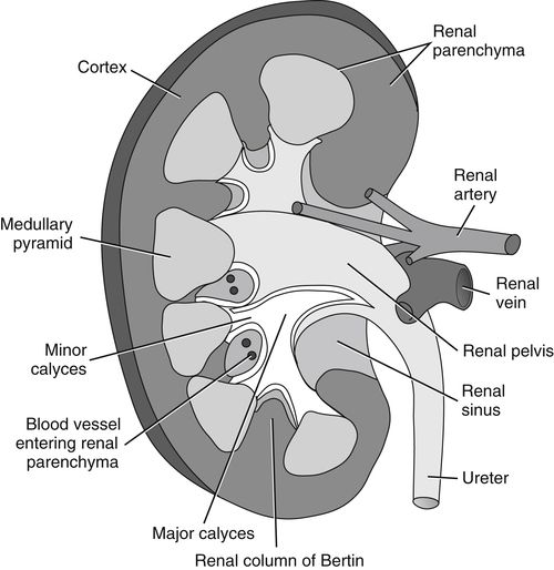
Figure 9-2 Section of internal kidney anatomy. Observe how the ureter begins in the kidney as the renal pelvis. Notice how the number of medullary pyramids equals the number of minor calyces. Note the columns of Bertin separating each medullary pyramid. B. Outer cortex: The outer renal cortex lies between the medulla and outer renal capsule. It contains millions of nephrons; the microscopic functional units of the kidney that form urine.PhysiologyAs part of the of the excretory system kidneys function to get rid of metabolic wastes and purify the blood by excreting urine (Excess water, salt, & toxins)next | |
| | | | د.كمال سيد
Admin
عدد المساهمات : 2679
نقاط : 4483
السٌّمعَة : 9
الجنس :
علم بلدك :
تاريخ الميلاد : 03/04/1950
تاريخ التسجيل : 30/07/2012
العمر : 74
الموقع : السودان - سنار
العمل/الترفيه : طبيب عمومى وموجات صوتية
الساعة الان :
دعائي :
| | موضوع: Renal Scanning Protocol contd أمس في 6:53 pm | |
| Renal Scanning Protocolcontd* As seen in the following transverse scanning plane image of an axial section of the midportion of the right kidney, 1.the overall sonographic appearance of the normal adult kidney is described : as heterogeneous (mixed echo pattern) due to the contrast in appearance of the renal cortex and renal sinus. The 2.renal cortex appears +midgray and +homogeneous (uniform echo pattern) with a +smooth contour whereas the 3.renal sinus presents as +bright ( hyperechoic) with +irregular borders and, in this case, +interrupted with +anechoic, urine-filled medullary pyramids and blood-filled vessel. Renal Scanning Protocol | Radiology Key | |
| | | | | | Abdominal Ultrasound | |
|
مواضيع مماثلة | |
|
| | صلاحيات هذا المنتدى: | لاتستطيع الرد على المواضيع في هذا المنتدى
| |
| |
| |
|