موقع د. كمال سيد الدراوي
طبي_ اكاديمي _ ثقافي _ تعليمي _ _ استشارات طبية_فيديو طبي
|
| | | مقدمة تعريفية عن الموجات فوق الصوتية |  |
| | | كاتب الموضوع | رسالة |
|---|
د.كمال سيد
Admin


عدد المساهمات : 2464
نقاط : 4252
السٌّمعَة : 9
الجنس : 
علم بلدك : 
تاريخ الميلاد : 03/04/1950
تاريخ التسجيل : 30/07/2012
العمر : 74
الموقع : السودان - سنار
العمل/الترفيه : طبيب عمومى وموجات صوتية
الساعة الان :
دعائي : 
|  موضوع: مقدمة تعريفية عن الموجات فوق الصوتية موضوع: مقدمة تعريفية عن الموجات فوق الصوتية  الثلاثاء سبتمبر 11, 2012 5:45 am الثلاثاء سبتمبر 11, 2012 5:45 am | |
| INTRODUCTION to ULTRASOUND
Dr. Kamal Sayed Ismaeel Eldirawi MsC diagnostic medical ultrasound AAU Khartoum Sudan - AAIMS Pakistan : الأشعة والموجات فوق الصوتية
ونبدا بتعريف الصوت : الصوت
الصوت عبارة عن موجات أو اهتزازات ميكانيكية تصل إلى الأذن ثم تتحول عبر الأذن إلى اهتزازات تصل إلى خلايا الدماغ حيث تتجاوب معها بل وتغير من اهتزازات خلايا الدماغ، تسير هذه الاهنزازات في الهواء بسرعة تبلغ 340 متراً في الثانيةتقريباً، ولكل صوت من الأصوات هناك تردد معين، .وتنتشر هذه الأمواج في الهواءثم تتلقّاها الأذن، ثم تنتقل عبر الأذن حيث تتحول إلى إشارات كهربائية وتسير عبرالعصب السمعي باتجاه اللحاء السمعي في الدماغ، وتتجاوب الخلايا معها ومن ثم تنتقلإلى مختلف مناطق الدماغ وخصوصاً المنطقة الأمامية منه، وتعمل هذه المناطق معاً علىالتجاوب مع الإشارات وتترجمها إلى لغة مفهومة للإنسان. وهكذا يقوم الدماغ بتحليلالإشارات ويعطي أوامره إلى مختلف أجزاء الجسم ليستجيب لهذهالإشارات. ولكل صوت من الأصوات هناك تردد معين، ويتراوح المجال المسموع للإنسان من 20 ذبذبة في الثانية إلى 20000 ذبذبة في الثانية ويمكن القول ان الصوت هو تردد آلي، أو موجة قادرة على التحرك في عدة أوساط مادية مثل الأجسام الصلبة، السوائل، و الغازات، ولاتنتشر في الفراغ , وباستطاعة الكائن الحي تحسسه عن طريق عضو خا ص يسمى الأذن. من منظور علم الأحياء فالصوت هو إشارة تحتوي على نغمة أو عدة نغمات تصدر من الكائن الحي الذي يملك العضو الباعث للصوت، تستعمل كوسيلة اتصال بينه وبين كائن آخر من جنسه أو من جنس آخر، يعبر من خلالها عما يريد قوله أو فعله بوعي أو بغير وعي مسبق، ويسمى الأحساس الذي تسببه تلك الذبذبات بحاسة السمع وتقدر سرعة الصوت في وسط هوائي عادي ب 340 متر في الثانية او 1026 كم في الساعة. و تتعلق سرعة الصوت بعامل الصلابة وكثافة المادة التي يتحرك فيها الصوت.الصوت هو اهتزاز ميكانيكي للوسط ، الصوت ليس موجة بل الموجة هي إحدى الاشكال (نماذج الانتشار) التي يبرز و يتميزبها الصوت و كمثال على نماذج اخرى: التيارات الصوتية و التدفق الصوتي هنالك عوامل اخرى تؤثر على انتشار الصوت وسرعته كطبيعة المادة (اللزوجة، تأثرها بالمجال المغناطيسي) سرعة الصوت
تختلف سرعة الصوت حسب نوع الوسط الذي تنتشر فيه الموجات الصوتية و درجة الحرارة فتكون أعلى في المواد الصلبة وأقل في السوائل وأقل بكثير في الغازات. وبالنسبة لانتشار الصوت في الهواء فيعتمد على الضغط ، أي أن سرعة الصوت تقل بالارتفاع عن سطح الأرض. فمثلا سرعة الصوت في الهواء عند درجة الصفر المئوي هي 331.1 م/ث وتزداد هذه السرعة بارتفاع درجة الحرارة. تقدر سرعة الصوت في الماء بـ1450 م/ث عند الدرجة القياسية (15 درجة مئوية). وتتراوح هذه السرعة في المواد الصلبة بين 3000 و 6000 متر/ثانية فهي مثلا 5100 م/ث للحديد والألمنيوم و3560 م/ث للنحاس وتبلغ 5200 متر في الثانية في الزجاج.[/size]
يعتبر الصوت أحد الظواهر الهامة التي يستعملها الإنسان والحيوان للتخطيط والتفاهم عن طريق حاسة السمع (الاذن) التي يتم بواستطها تحويل الصوت من موجات صوتية إلى أشارات كهربائية عن طريق الاذن والمخ والتي تتحول إلى معلومات مفهومة وتشمل هذه الظواهر جميع الاصوات على اختلاف مصادرها ووسائلها.مثلا سماع الاصوات من الآلات الموسيقية و تعدد وسائل الاتصالات المسموعة التي تعتمد على تحويل الطاقة من صورة إلى أخرى وتطور الأجهزة الصوتية التي تأخد أشكالا متعددة في تطبيقاتها الحديثة في مجالات الطب والصناعة والزراعة وغيرها تجعل العلماء والمهتمين بهذا المجال يكثفون الجهد لفهم الظواهر الموجية من حيث مصادرها وكيفية حدوثها وطرق انتشارها والعوامل التي تتحكم فيها ومدى الاستفادة منها. إذا لاحظنا بعناية الطرق التي يحدث بها الصوت نجد أنه لابد من بدل شغل في كل حالة.الموسيقى يبذل شغلا لتحريك أوتار الآلة الموسيقية كما أن الصوت الناتج عندما تصفق يديك لتشجيع فريقا رياضيا مثلا يأتي من بذل شغل وهذا الشغل المبذول بواسطة اليدين يسبب اضطرابا في الهواء المحيط منحولا إلى طاقة صوتية تتشكل على شكل موجات منتظمة عليه فإن الصوت صورة من صور الطاقة إذا استقبلتها الأذن يحدث الاحساس بالسمع.و تعتبر دراسة "الصوت" من المواضيع المهمة حيث تستخدم هذه الدراسات في ابحاث الطيران والفضاء والطاقة المتجددة والطاقة النووية والابحاث الطبية. و يمكن توليد الصوت بوسائل ميكانيكية أو حرارية. وتستخدم الوسائل الحرارية في بناء المبردات الصوتية الحرارية وكذلك في عمليات الكشف عن الماء الموجود في النفط السماع عند الحيوانات
تختلف درجة السماع في الحيوانات من فصيلة لأخرى. الخفاش : يقوم بتوليد موجات فوق صوتية تصل إلى 100 كيلوهرتز ثم الاستماع إليها لتكوين صورة للاجسام المحيطة به. الكلب: يستطيع سماع الاصوات الأعلى ترددا من السمع البشري. الأسماك: بعض الاسماك تستمع إلى اصوات بترددات تصل إلى 180 كيلوهرتز والبعض الاخر إلى 4 كيلوهرتز فقط الاذن البشرية
تستطيع الأذن البشرية سماع الاصوات التي تتراوح تردداتها بين 20 هرتز و 20 كيلوهرتز بوضوح بينما تتوهن جميع الاصوات التي تقل عن 20 هرتز أو تزيد عن 20 كيلوهرتز (20.000 هرتز). لهذا السبب يقوم مصمموا السمعيات بوضع ما يسمى مرشح إلكتروني لتمرير الترددات السمعية فقط كما هو الحال في بطاقات الصوت الخاصة بالحواسيب مثلا.  يتبع
عدل سابقا من قبل د.كمال سيد في الإثنين أبريل 22, 2019 9:44 pm عدل 4 مرات | |
| | | | د.كمال سيد
Admin
عدد المساهمات : 2464
نقاط : 4252
السٌّمعَة : 9
الجنس :
علم بلدك :
تاريخ الميلاد : 03/04/1950
تاريخ التسجيل : 30/07/2012
العمر : 74
الموقع : السودان - سنار
العمل/الترفيه : طبيب عمومى وموجات صوتية
الساعة الان :
دعائي :
| | موضوع: رد: مقدمة تعريفية عن الموجات فوق الصوتية الثلاثاء سبتمبر 11, 2012 6:17 am | |
| تصنيف الصوت تبعا للتردد
بحسب التردد يصنف الصوت إلى الأنواع :
• تحت الصوتية ، وهي أقل من 16 هرتز وهي غير مسموعة للأذن البشرية حيث التردد منخفض جدا ،
• نطاق السمع (audible range) , وهو يمتد من 16 هرتز إلى نحو 20.000 هرتز ، وهي أصوات مسموعة للبشر ،
• فوق صوتية ، بين 20.000 هرتز إلى 6و1 جيجا هرتز (6و1 مليار ذبذبة في
• الثانية) ، وهي غير مسموعة للبشر ، حيث ترددها عالي.
(ويمكن ان يكون هذا اجابة للسؤال : ما معنى الموجات فوق الصوتية ؟).
• تصواتي أو فوق صوتي (بالإنكليزية: Ultrasonic or Ultrasound) مصطلح يطلق على الترددات الصوتية التي تفوق 20 كيلوهرتز. القيمة 20 كيلوهرتز هي قيمة تقريبية وتختلف من أذن بشرية لأخرى.
تصنيفات الموجات الصوتية
تصنف الموجات الصوتية طبقا لتردداتها كما يلي:
الموجات المسموعة AUDIBLE WAVES
هي تلك الموجات التي تقع تردداتها بين 20 هرتز و 20.000 هرتز ، وتمثل الصوت المسموع بواسطة الأذن البشرية العادية. حيث أن الحد الأدنى لتردد الصوت التي تحس بها الأذن البشرية الطبيعية هو 20 هيرتز تقريبا بينما الحد الأعلى هو 20 الف هرتز ، وينخفض هذا المدى عند كبار السن إلى حوالي 12.000 هرتز. وأقصى درجات الاحساس بالصوت لأذن بشرية عادية يقع في المدى بين 5000 هيرتز و8000 هيرتز والذي يشمل ذبذبات الحروف الهجائية. وكما هو معروف يمكن أحداث الموجات السمعية عن طريق الاحبال الصوتية في الإنسان والآلات الموسيقية سواء الوترية أو النحاسية أو الأنبوبية وغيرها من الآلات الأخرى.
الموجات الفوق سمعية ULTRASOUND WAVES
هي الموجات التي تزيد تردداتها على 20 الف هيرتز والتي تقع خارج نطاق حاسة الاذن البشرية. وهذا النوع من الموجات ما زال موضع بحث واهتمام مكثف نظرا للتطبيقات المهمة التي تمس مجالات عديدة في الصناعة والطب وغيرهما. وقد أصبح بالإمكان إنتاج موجات فوق صوتية تزيد تردداتها على 1000000 هيرتز ولاتختلف هذه الموجات من حيث الخواص عن الموجات الصوتية الاخــرى إلا أنه نظرا لقصر طول موجاتها فإنه بالإمكان تنتقل على هيئة أشعة دقيقة عالية الطاقة.
الموجات تحت السمعية INFRASOUND
هي الموجات الصوتية التي يقل ترددها عن 20 هيرتز ولاتستطيع الاذن البشرية الاحساس بها واهم مصدر لها هو الحركة الاهتزازية والانزلاقية لطبقات القشرة الأرضية وما ينتج عنها من زلازل وبراكين وعليه انها مهمة جدا في رصد الزلازل وتتبع نشاط البراكين. وتستطيع بعض الحيونات الاحساس بالزلازل قبل حدوثها بسببها
تطبيقات الترددات التصواتية (فوق الصوتية)
يمكن تصميم مولدات فوق صوتية وأجهزة تحسس فوق صوتية لاستخدامها في الكثير من التطبيقات الصناعية والطبية مثل:
• السونار (رادار فوق صوتي بنطاق عريض يسمح بتصوير ثلاثي البعد)
• الاشعة التلفزيونية الثنائية والثلاثية البعد.
• قتل بعض أنواع البكتيريا
• المنظفات فوق الصوتية
موجات طولية وموجات عرضيةا
عدد من موجات جيبية ذات ترددات مختلفة ; الموجات السفلى لها تردد أعلى من الموجات العليا في الشكل. المحور الأفقي يمثل الزمن.
ينتشر الصوت في الغازات والبلازما وفي السوائل على هيئة موجات طولية ، وتسمى عند الفيزيائيين موجات ضغطية. أما في المواد الصلبة فينتشر الصوت فيها كموجات طولية وأيضا موجات عرضية. وتتكون موجات الصوت الطولية من تتابع لطبقات يعلو فيها الضغط وطبقات يقل فيها الضغط عن الضغط المتوازن المعتاد متتابعة. أما الموجات العرضية في المواد الصلبة فهي موجات متتابعة من إجهاد جزي عرضي ، يكون عموديا على اتجاه انتشار الصوت.
وفي موجات الصوت تنزاح جزيئات الوسط دوريا وتهتز ، ولكنها لا تنتقل مع الصوت. وتنتقل الطاقة المحمولة مع الصوت كطاقة حركة لاهتزازات الوسط.
التعريف الفيزيائي
من وجهة نظر الفيزياء فالصوت هو موجة. وتكون الموجة في السوائل والغازات موجة طولية وهي كذلك أيضا في الهواء. أما في المواد الصلبة فينتشر الصوت في موجات عرضية. وتحرك الموجات جزيئات الوسط (غالبا الهواء) حول حالة وسطية وتنتشر بسرعة خاصة ، ويرمز لسرعة الصوت c.وتنقل الموجات طاقة صوتية. ولا ينتشر الصوت في الفراغ.
وتعتمد سرعة الصوت على الوسط الذي ينقلها. وتبلغ سرعة الصوت في الهواء 343 متر في الثانية عند درجة حرارة 20 درجة مئوية و 1407 متر /ثانية في الماء عند درجة الصفر المئوي.
يمكن حساب طول الموجة الصوتية λ من تردد الموجة f وسرعة الصوت c بواسطة المعادلة:
وفي العادة تكون اختلافات في الضغط أو في الكثافة سببا في تغير سرعتها. ويتضح هذا عندما نتصور مستوي لضغط الصوت يقدر ب 130 dB ديسيبل : وهذا يبلغ درجة تألم أذن الإنسان ، ويمثل به الضغط الجوي العادي : يبلغ الضغط الجوي للهواء الساكن 101325 باسكال ، في حين أن مستوي ضغط صوت قدره 130 dB له قيمة فعلية لضغط الصوت p تبلغ 63 باسكال فقط.
اسرع من الصوت (خارق الصوت TRANS SONIC)
يستخدم مصطلح "خارق صوت" للإشارة للسرعة التي تزيد عن سرعة الصوت (1 ماخ). في درجة حرارة 21 مئوية (70 فهرنهايت) تعد القيمة المطلوبة بداية لأي جسم ليتحرك بسرعة خارقة صوت هي تقريبا 344 متر في الثانية (1.129 قدم/ثانية أو 761 ميل في الساعة أو 1,238 كيلومتر في الساعة).
يطلق أحيانا مصطلح هايبر سونيك على السرعات التي تزيد 5 مرات عن سرعة الصوت.
تسمى السرعات التي يكون بعض أجزاء الهواء حول الجسم فيها (مثل نهاية شفرات المحرك الدوار) تصل لسرعة خارقة الصوت ترانس سونيك (تقريبا ما بين 0.8 الى 1.2 ماخ).
| |
| | | | د.كمال سيد
Admin
عدد المساهمات : 2464
نقاط : 4252
السٌّمعَة : 9
الجنس :
علم بلدك :
تاريخ الميلاد : 03/04/1950
تاريخ التسجيل : 30/07/2012
العمر : 74
الموقع : السودان - سنار
العمل/الترفيه : طبيب عمومى وموجات صوتية
الساعة الان :
دعائي :
| | موضوع: رد: مقدمة تعريفية عن الموجات فوق الصوتية الثلاثاء سبتمبر 11, 2012 6:33 am | |
| مصطلحات خاصة بعلوم الموجات فوق الصوتية
اليكم بعض المصطلحات التى تعترض طريقنا فى فهم اساسيات الموجات الصوتية --- ارجو الصبر عليها
Acceleration - increase in velocity per unit time.
Aneurysm - a sac formed by the localized dilatation of the wall of an artery, vein, or the
heart.
Atherosclerosis - a common form of arteriosclerosis in which deposits of yellowing plaques
(atheromas) containing cholesterol, other lipoid material, and lipophages are formed within
the intima of large and medium-sized arteries. The word atherosclerosis comes form the
Greek, athere, meaning “soft, fatty, gruel-like,” and scler- meaning “hard”. These terms are
descriptive of the material deposited on the intima of an artery and of the state of the
arterial muscle walls once they have been affected by the disease.
Bernoulli effect - pressure reduction in a region of high flow velocity.
Compliance - dispensability; nonrigid stretchability of vessels. adj. compliant -
Energy - power that may be translated in motion (kinetic), overcoming resistance, or
effecting physical change; the ability to do work. Energy assumes several forms; it may be
thermal (in the form of heat), electrical, mechanical, chemical, radiant, or kinetic. In doing
work, the energy is changed from one form to another or to several forms. In these
changes some of the energy is “lost” in the sense that it cannot be recaptured and used
again. Usually there is loss in the form of heat, which escapes or is dissipated unused. All
energy changes give off a certain amount of the energy as heat.
Flow - the movement of a liquid or gas.
Flow resistance - pressure difference divided by volume flow rate for steady flow.
Friction - the force that resists the movement of one object on another.
inertia - resistance to acceleration.
Intima - the innermost lining of a blood vessel. Also called tunica intima.
laminar flow - flow in which fluid layers slide over each other in a smooth, orderly manner,
with no mixing between layers.
Poiseuille’s law - the mathematical description of the dependence of volume flow rate on
pressure, vessel length, radius, and fluid viscosity.
Pressure - force divided by area in a fluid. blood p. - the pressure of the blood on the walls
of the arteries, dependent on the energy of the heart action, elasticity of the arterial walls,
and volume and viscosity of the blood;
The maximum or systolic pressure occurs near the
end of the stroke output of the left ventricle, and the minimum or diastolic late in ventricular
diastole.
Pulse pressure. - the difference between the systolic and diastolic blood pressures.
Pulsatile flow - flow that accelerates and decelerates with each cardiac cycle.
Stenosis - a narrowing of the lumen of a blood vessel, most commonly due to
atherosclerosis.
Turbulent flow - also turbulence; random, chaotic, multidirectional flow of a fluid with
mixing between layers; nonlaminar flow.
Velocity (linear) - refers to the rate of displacement of a particle with respect to time. The
units used to describe velocity are distance per unit time e.g. cm/sec.
Viscosity - resistance of a fluid to flow; a physical property of a fluid that is dependent on
the friction of the component molecules as they slide by one another. adj. viscous - having
a high degree of viscosity.
Volume flow rate - the volume of a fluid that passes through an organ or part in a specified
time. Expressed in units of volume per unit time, i.e. cm /sec.
يتبع
| |
| | | | د.كمال سيد
Admin
عدد المساهمات : 2464
نقاط : 4252
السٌّمعَة : 9
الجنس :
علم بلدك :
تاريخ الميلاد : 03/04/1950
تاريخ التسجيل : 30/07/2012
العمر : 74
الموقع : السودان - سنار
العمل/الترفيه : طبيب عمومى وموجات صوتية
الساعة الان :
دعائي :
| | موضوع: رد: مقدمة تعريفية عن الموجات فوق الصوتية الثلاثاء سبتمبر 11, 2012 6:45 am | |
| MORE TERMINOLOGY
TERMINOLOGY ASSOCIATED WITH IMAGE CHARACTERISTICS
The accurate description and interpretation of sonograms depends on the correct use of related terms associated with the reflection and attenuation characteristics of structures.
Masses are categorized a solid, cystic, or complex depending on the observed sonographic characteristics
Terms Related To Echogenicity
Structures with acoustic ( means pertaining to sound waves) interfaces show different degrees of reflectivity which translates into different levels of echogenicity in the sonogram.
Echogenic: a region in an ultrasound image which has echoes. Synonymous terms: reflective, echo-producing, echoic.
Anechoic: literally means “without echoes”. An anechoic region in an ultrasound
image contains no echoes. Synonymous term: echo-free. Another term that is erroneously used is “echolucent”. This term has come from Radiology where the term radiolucent is used to describe dark-appearing areas on a radiograph representing tissues that permit passage of x-rays. Use the term anechoic rather than the term echolucent.
“Transonic” is not a synonymous term although sometimes it is inappropriately used to mean anechoic. Transonic describes an area of relatively low attenuation. As an example, the normal gallbladder is transonic therefore acoustic enhancement is seen distal to it. If the gallbladder contains 'sludge' (echogenic bile) it has real echoes in it (it is no longer anechoic) but it remains transonic relative to soft tissue and still produces acoustic enhancement.
Hyperechoic: describes a structure which has many echoes. Synonymous term : hyperechogenic.
Hypoechoic: in an ultrasound image, describes a structure which has few echoes. Synonymous terms: echo poor, echopenic .
Isoechoic: having the same echo density or brightness as another structure.
As an example, the pancreas may be isoechoic to the liver.
If a solid lesion is isoechoic to the surrounding parenchyma, it may not be visualized unless it has a displacement or contour effect. The better a system’s contrast resolution, the better the ability to distinguish between normal and abnormal tissues with similar echo amplitudes.
Homogeneity, Heterogeneity :
Homogeneity (adj. homogeneous) and heterogeneity (adj. heterogeneous) are terms relating to the histologic make up of normal and abnormal tissues in relationship to their echogenic characteristics.
Homogeneous structures are histologically uniform throughout whereas heterogenous structures are non uniform.
Solid masses are generally echogenic, with variable levels of echo amplitudes.
Homogeneous solid masses have few reflectors and will appear to be relatively
echo poor on the display. Conversely, solid masses that are heterogeneous contain numerous reflectors and will appear echogenic (variable levels of echogenicity dependent on several factors).
The terms homogeneous and heterogeneous may also be used to describe the
echogenic appearance of structures.
Structures that are homogeneous display a uniform echo pattern and the tissue texture will have a uniform dot size, distribution, and brightness. A good example of a normal structure with a homogeneous appearance is the spleen .
Conversely, structures that are heterogeneous display a non-uniform echo pattern. Liver with metastases frequently has a heterogeneous echo appearance .
Solid, Cystic, and Complex Structures :
Scanned structures may be described as being solid, cystic or complex when displayed depending on their sonographic appearances.
Solid structures are composed of tissue and are associated with variable levels of echogenicity. They may be either homogeneous or heterogeneous, and exhibit variable degrees of attenuation. In comparison to cystic structures, solid structures are echogenic and are associated with average to above average attenuation. The testes and liver are two examples of solid structures.
Cystic structures contain fluid. In comparison to solid structures, cystic structures
are anechoic, and associated with decreased attenuation and the display of acoustic enhancement. Simple cysts are anechoic, have a thin well-defined walls, and produce good acoustic enhancement.
Complex cysts are cysts that feature one or more of the following characteristics : echogenic fluid (diffuse or layered), septations, or a thick or irregular wall . Complex masses contain both solid tissue and fluid.
Texture is the echo pattern (dot size, distribution and brightness) generated by tissues. The displayed echo texture depends on several technical factors including transducer frequency, dynamic range, output power, gain, and pre- and postprocessing algorithms.
Speckle is the displayed dot pattern of organ parenchyma due to constructive and destructive wave interference from adjacent scatterers. The result of this acoustic interference produces a display dot pattern that does not represent the individual scatterers in the tissue but rather represents the resultant interference pattern from all the scatterers within the region.
Speckle is considered a type of acoustic artifact which degrades tissue echo texture. Speckle tends to produce a very fine dot pattern in the near field which become coarser in the far field because attenuation filters out the higher frequency echoes.
The sonographer can not correct acoustic speckle (only recognize it for what it is). Since speckle is not a favorable acoustic event, equipment manufacturers continue to work to reduce it.
يتبع
عدل سابقا من قبل د.كمال سيد في الجمعة سبتمبر 14, 2012 8:22 pm عدل 1 مرات | |
| | | | د.كمال سيد
Admin
عدد المساهمات : 2464
نقاط : 4252
السٌّمعَة : 9
الجنس :
علم بلدك :
تاريخ الميلاد : 03/04/1950
تاريخ التسجيل : 30/07/2012
العمر : 74
الموقع : السودان - سنار
العمل/الترفيه : طبيب عمومى وموجات صوتية
الساعة الان :
دعائي :
| | موضوع: رد: مقدمة تعريفية عن الموجات فوق الصوتية الثلاثاء سبتمبر 11, 2012 6:57 am | |
|
نبذة تاريخية عن الموجات فوق الصوتية
كانت أولى مساعي البحث في الموجات الصوتية منذ عام 1822 عندما سعى عالم الفيزياء السويسري ( دانيل كولادين ) لحساب سرعة الصوت عن طريق جرسه المائي في مياه بحيرة (جنيفا ) . والتي مهدت لوضع ( نظرية الصوت ) في عام 1877 بجهود العالم ( لورد ريليه ) والتي شرحت الأساسيات الفيزيائية لموجات الصوت وانتقاله وارتداده . وتوالت الأبحاث حتى كان تصميم وانشاء أول نظام( Sound Navigation & Ranging ) Sonar عامل في الولايات المتحدة عام 1914 على يد العالم فسندن لأغراض الملاحة البحرية ولتحديد أماكن المارينز الألماني في الحرب العالمية الأولى.
ولم توظف الموجات فوق الصوتية لخدمة الأغراض الطبية حتى بداية الأربعينات على يد دكتور الأعصاب والطبيب النفسي النمساوي ( كارل ثيودور ) والذي يعتبر أول طبيب استخدم الموجات فوق الصوتية في التشخيص الطبي وقد واجه في ذلك صعوبات بسبب امتصاص عظام الجمجمة لمعظم طاقة الموجات فوق الصوتية.
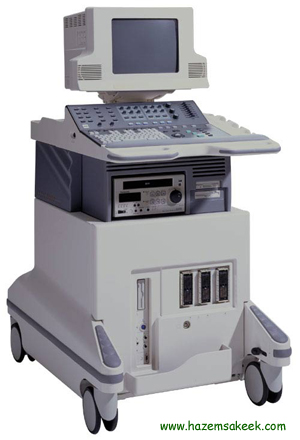
وبعد حصيلة جهود مكثفة للفيزيائيين والمهندسين الميكانيكيين والكهربائيين والبيولوجيين بالتعاون مع الأطباء ومبرمجي الكمبيوتر والباحثين ودعم الحكومات,ابتدأ التشخيص بالموجات فوق الصوتية ليأخذ محله في عيادات الأعصاب والقلب والعيون ولتتطور الموجات منA-Mode محدودة الاستخدام الىB-Modeوالتي سعى العالم ( دوغلاس هوري ) كفني أشعة لاستغلالها في التشخيص لقدرتها على اختراق الأنسجة بهدف الدراسة التشريحية لأعضاء الجسم في جامعة ( كولورادو) في دنغر بالتعاون مع زميله أخصائي الكلى ( جوزيف هوملس ) والذي بدوره تبنى الأبحاث الطبية على هذا الصعيد وقام بتوجيهها وبتعاون العلماء والمهندسين ( بيلز و بوساكوني ) كان أول جهاز ألتراساوند ثنائي الأبعاد يعمل بنظامB-Modeعام 1951
وتوالت الأجهزة التي تعمل في هذا النظام الا أنها جميعا كانت كبيرة الحجم وعلى المريض أن ينغمس كليا أو جزئيا في الماء في وضعية السكون لفترة زمنية طويلة الأمر الذي جعله غير عملي ويستحيل وجوده في عيادات الاختصاص.
الذراع المعدني المتحرك
وفي أواخر عام 1955 بدأ العالم بتطوير هذه الأجهزة لتصبح أكثر حساسية وأقل حجما وأكثر سهولة في طريقة الفحص حتى توصلوا للذراع المعدني المتحرك والذي يوضع على المكان المخصص للفحص , كما تم تطوير جهاز الألتراساوند المهبلي و الشرجي عام 1955 والذي يعمل بنظامA-Mode بجهود العالمين ( وايلد ) و ( ريد ) الا أنه في هذه المرحلة لم يحقق الغرض الفعلي حتى تم تطويره لاحقا , كل هذه التطورات والتي مرت بنجاح باهر وأخذت محلها في جزء كبير من العالم كالولايات المتحدة الأمريكية, والنمسا, واليابان, والمملكة المتحدة , مهدت الطريق وفتحت المجال لدخول جهاز الالتراساوند إلى تخصص النسائية والتوليد وهكذا كانت الانطلاقة.
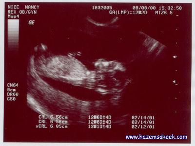
ومن رواد تسخير الالتراساوند لخدمة هذا التخصص الدكتور الانجليزي( ايان دونالد ) والذي له مساعي رائدة على هذا الصعيد حيث ابتدأ طريقه بتشخيص التكتلات البطنية سواء أكانت ألياف أو أورام أو حتى أكياس وخرج بورقة عمل كانت من أعظم الاشراقات الطبية خلال العشرة أعوام المنصرمة بتعاونه مع العالم الفني ( توم براون ) والدكتور ( جون ماكفيكار ) في 7 / 6 / 1958 وعام 1959 استطاع أن يلتقط أصداء واضحة لرأس الجنين ومن بعدها أصبحت أبعاد محيط رأس الجنين هي الوسيلة المعتمدة لدراسة نمو الجنين . و بعد مرور سنوات قليلة كان بالامكان دراسة الحمل منذ البداية حتى النهاية , وتشخيص كثيرا من المشاكل كتعدد التوائم والتشوهات الخلقية والمشاكل التي تصاحب المشيمة.
ولم يكن قبل عام 1972 بالامكان رؤية ودراسة الحويصلات بنظامB-Mode على يد العالم النمساوي( Kratochwil )وهكذا أصبح التصوير بالموجات فوق الصوتية في مجال الأمراض النسائية والتوليد مضمارا للتنافس بين الاختصاصيين وتزايدت الأبحاث وأوراق البحث من ورقة بحث واحدة للدكتور (ايان دونالد) عام 1958الى 296 ورقة عمل في عام 1978.
وبهذه الجهود المكثفة استطاع الأطباء تشخيص كيس حمل بعمر 5 أسابيع في عام 1963 , وتحديد نبض الجنين بعمر 7 أسابيع عام 1965وفي السبعينات أصبح بالإمكان قياس محيط رأس الجنين ومحيط صدره والتي لعبت دورا جوهريا في متابعة نمو الجنين وتطوره واكتشاف أي إعاقة في النمو, وحساب طول الجنين ومحيط البطن الذي استطاع العلماء من خلاله أخذ فكرة عن وزن الجنين وظروف تغذيته.
كما تم تشخيص فتحات الظهر ( Spina Bifida )واختفاء جمجمة الرأس في الأجنة( Anencephaly )في الأسبوع السابع عشر من الحمل. كل تلك التطورات لم تكن لتكون لولا إيجادB-Modeودخول درجات اللون الرمادي على أجهزة الالتراساوند بعد أن كانت في اللون الأبيض والأسود, وهذه الدرجية في اللون أعطت وضوح في الصورة وأصبح تركيز العلماء على زيادة هذه الدرجية لزيادة الدقة في الفحص.
ومع الثمانينات حدثت ثورة حقيقية في عالم الموجات فوق الصوتية وهي ما يسمى( Real time scanner )أي التصوير الحي ( ثنائي الأبعادB-Mode ) والذي عن طريقه تم التعرف على حياة الجنين الفعلية, وحركاته, وتصرفاته, ونبضات القلب, والتنفس في رحم الأم. وكان أول جهاز فعال في هذا المجال عام 1985 في ألمانيا , وكانت الثمانينات هي ميدان التنافس للشركات المصنعة لأجهزة الالتراساوند لتقديم أدق الصور وأوضحها. وهكذا اتضحت معالم علم جديد في تخصص النسائية والتوليد ( تشخيص وسلامة الجنين) .
كما تم تطوير جهاز الالتراساوند المهبلي والشرجي , والذي صمم لأول مرة على يد( ريد ) و( وايلد ) عام 1955, وقد أستغرق هذا التطوير حوالي العشرين عاما ليصبح فعالا وليحقق طموحات العلماء في كشف الأعضاء الداخلية للحوض ولينتشر بين الأوساط الطبية.
وكان عام 1985 هو العام الذي احتضن أكثر أجهزة الالتراساوند المهبلي فعالية وأعظمها فائدة ,وقد تزامن تطويره مع أولى تقنيات أطفال الأنابيب في النمسا.
ومن التقنيات التي استغرقت زمنا طويلا حتى سخرت للاستعمال الفعلي الدوبلار( ( Doppler و( M-Mode ) فمبدأ الدوبلار كان أول وصف وضع له بجهود العالم النمساوي ( كريستيان دوبلار ) عام 1842 وبدأ بتطبيقه اليابانيون عام 1955 لدراسة حركات وصمامات القلب. وفي عام 1962 وباستخدام الدوبلار تتبع العلماء تدفق الدم ونبضات الجنين وتطورت الأجهزة في تقصي التدفقات الدموية لتصبح ثنائية الأبعاد وتمكن من المتابعة الحية الملونة لسير الدم(( Real time Color Flow Imaging .
وكانت الثمانينات ومطلع التسعينات مسرحا للتقدم العظيم الذي أحرزته الشركات المصنعة لأجهزة الالتراساوند ثنائية الأبعاد كما أصبحت تلك الأجهزة عماد التأسيس لأي عيادة نسائية وتوليد , وساعد في ذلك الحجم المعقول وأذرعة الفحص والتي تشغل حيزا ضيقا على الجسم وتعطي مجالا واسعا للرؤية إضافة إلى الحركة الحرة لهذه الأجهزة.
والأهم من كل ذلك التفاصيل الدقيقة التي تمحصها تلك الأجهزة بوضوح الصورة وتحديد الملامح والتشخيص الدقيق بحيث احتلت أجهزة الالتراساوند المرتبة الأولى من بين وسائل التشخيص, والتي لم تقتصر على تشخيص الجسم فحسب بل تجاوزتها لفحص الأجهزة الداخلية للأجنة في أرحام أمهاتهم لتصبح علما بل وتخصصا قائما بذاته.
وبعد هذه المراحل العريقة في تاريخ الموجات فوق الصوتية وبعد ثورات العلم المتأججة على كل صعيد ومتطلبات العصر المتجددة مع دنوّ الألفية الثانية وارتباط الكمبيوتر الوثيق بكل التحركات البشرية مهما صغرت , غدت أجهزة الالتراساوند الثنائية الأبعاد غير مرضية- بالرغم من كل النجاح الذي حققته- وشخصت أعين العلماء نحو البعد الثالث الذي عجزت الأجهزة ثنائية الأبعاد عن سبر غوره وان كانت الفكرة تلوح في الأفق منذ السبعينات , الا أنها بدأت تتمحور وتأخذ أبعادها مع مطلع الثمانينات وأعظم ما ساند وجودها الثورات التكنولوجية في برمجة الكمبيوتر.
وفي اليابان في جامعة طوكيو كان أول تقرير حول نظام الأبعاد الثلاثية ( الطول, العرض, العمق أو الارتفاع ) عام 1984 وأول محاولة ناجحة في الحصول على صورة جنين ثلاثية الأبعاد من صورة ثنائية الأبعاد عن طريق الكمبيوتر كانت عام 1986.
وبعد تطوير أجهزة التراساوند مستقلة ثلاثية الأبعاد كانت المشكلة في الفترة الزمنية التي يستغرقها التقاط كل مقطع حيث تتجاوز العشر دقائق وهو ما يستحيل معه العمل سواء للطبيب المعالج أو المريض وبالتالي يستحيل معه التسويق. ومع الجهود المكثفة والتطوير المستمر كان أول جهاز التراساوند ثلاثي الأبعاد يأخذ محلا تجاريا في الأسواق في عام 1989Combison-330 ) )في النمسا واستمر العالم وخصوصا في اليابان, والنمسا, وبريطانيا, وكندا وحتى الصين في دفع عجلة التطور هذه حتى بدأت الأبحاث حول رباعي الأبعاد في لندن عام 1996 عندما بزغت فكرة التصوير ثلاثي الأبعاد الحي وليكون للبعد الرابع وهو البعد الزمني , دوره في إعطاء صورة حقيقية حيّة بأسلوب عملي , وما كان ذلك ليكون لولا التطورات الهائلة في علم الكمبيوتر والسرعة الهائلة في إجراء العمليات الحاسوبية
, ومن هنا كانت قصة البداية.
| |
| | | | د.كمال سيد
Admin
عدد المساهمات : 2464
نقاط : 4252
السٌّمعَة : 9
الجنس :
علم بلدك :
تاريخ الميلاد : 03/04/1950
تاريخ التسجيل : 30/07/2012
العمر : 74
الموقع : السودان - سنار
العمل/الترفيه : طبيب عمومى وموجات صوتية
الساعة الان :
دعائي :
| | موضوع: رد: مقدمة تعريفية عن الموجات فوق الصوتية الإثنين أبريل 22, 2019 9:43 pm | |
|
عدل سابقا من قبل د.كمال سيد في الإثنين مارس 25, 2024 6:06 pm عدل 2 مرات | |
| | | | د.كمال سيد
Admin
عدد المساهمات : 2464
نقاط : 4252
السٌّمعَة : 9
الجنس :
علم بلدك :
تاريخ الميلاد : 03/04/1950
تاريخ التسجيل : 30/07/2012
العمر : 74
الموقع : السودان - سنار
العمل/الترفيه : طبيب عمومى وموجات صوتية
الساعة الان :
دعائي :
| | موضوع: رد: مقدمة تعريفية عن الموجات فوق الصوتية الإثنين أبريل 22, 2019 9:45 pm | |
| What is Ultrasound:
. (ULTRASOUND is sound with a frequency too high for the human ear to hear (over 20 KHz
ECHOLOCATION is the ability of certain animals to produce pulses of sound (either audible or ultrasonic) and then to receive the returning echoes which are processed by the brain to give information about prey or obstacles.
The best known animal echolocator is of course the bat. There are more than 600 species of bats who use ultrasound in order to catch flying insects at night and to avoid obstacles. By rapidly vibrating their vocal cords they can emit short pulses of ultrasound at a frequency of up to 120KHz. These beams of ultrasound are beamed directionally into the darkness by means of the lips or noseflaps. If a moth happens to be cruising by, the sound waves are reflected from its body and return to the bat’s ears as echoes. When his brain has processed this information, he knows the size of the insect and its speed and direction and thus can swoop in on his prey in the darkness with great accuracy.
Just as a bat “sees” in the dark by emitting pulses of soundwaves, so the sonographer (ultrasound technologist) can “see” our insides by aiming high-frequency soundwaves, produced by a crystal with very special properties. There are a few differences of course. The frequencies used in medical ultrasound are much higher (from 1 to 5 MHz) whereas the maximum frequency for bats is 120 KHz.Another major difference is that the bat uses one organ to send sound (the vocal cords) and another to receive the echoes (the ears). In diagnostic ultrasonography the same crystal in the transducer both emits soundwaves andreceives the returning echoes as well.
Sound is made up of several different frequency waves. The very high frequency range is inaudible to the human ear and is known as ultrasound. Ultrasound was used by the Navy during World War II to detect submarines, and is widely used by fisherman to help find schools of fish.
In each case, an ultrasound machine is used. With the help of a microphone-shaped device (known as a transducer) ultrasound waves are created and beamed through water. When the beam encounters a boundary or interface between liquid (water) and a solid (submarine or fish) with a different density or compactness, part of the beam is reflected back to the transducer.
The remaining waves move through the object and reach the back boundary between solid and water. Here, some more of the ultrasound waves are reflected back to the transducer. In other words, the transducer transmits ultrasound and constantly receives waves that are reflected back every time the beam travels from one density to another.
The reflected ultrasound waves are collected and analyzed by the machine. Determining the amount of time it took for the beam to travel from and to the transducer (plus the the consistency and changes in position of the different structures that it passed through), the ultrasound machine can determine the shape, size, density and movement of all objects that lay in the path of the ultrasound beam.
The information is presented "real time" on a monitor screen and can also be printed on paper or recorded on tape, a CD or a computer disk. That is how warships detect submarines, fishermen identify choice fishing spots, an obstetrician evaluates the fetus of a pregnant woman, and a cardiologist examines the heart of a patient | |
| | | | د.كمال سيد
Admin
عدد المساهمات : 2464
نقاط : 4252
السٌّمعَة : 9
الجنس :
علم بلدك :
تاريخ الميلاد : 03/04/1950
تاريخ التسجيل : 30/07/2012
العمر : 74
الموقع : السودان - سنار
العمل/الترفيه : طبيب عمومى وموجات صوتية
الساعة الان :
دعائي :
| | موضوع: رد: مقدمة تعريفية عن الموجات فوق الصوتية الإثنين أبريل 22, 2019 9:45 pm | |
| [size=34]ما أهمية السونار للمرأة الحامل؟
يعتبر السونار من صور التكنولوجيا الحديثة الآمنة على الأم الحامل و جنينها ، متى يستخدم و ما هي نوع المعلومات المستقاه من السونار اكتشفي معنا فحص الموجات الصوتية لتصوير الجنين هو واحد من تقنيات التصوير التي تستخدم موجات صوتية عالية التردد لتكوين صور عن الجنين داخل الرحم. تساعد هذه الصور في تقييم نمو وسلامة الجنين ومتابعة أحداث تطور الحمل. كما يستخدم هذا الفحص أحيانا لاكتشاف بعض الحالات المرضية والتشوهات الخلقية التي تصاب بها الأجنة. يتم إجراء أول فحص بالموجات الصوتية غالبا في أشهر الحمل الأولى غالبا لتأكيد حدوث الحمل وتوثيق تاريخه، ومرة ثانية ما بين الأسبوع الـ18 و الـ 20 حين تصبح الصفات التشريحية للجنين أوضح، ويمكن تكراره بحسب الحاجة طوال مدة الحمل. [rtl]http://www.dailymedicalinfo.com/index.p ... eshow/s-10[/rtl] الرحلة : من البويضة إلى تكوين الجنين [rtl]http://www.dailymedicalinfo.com/index.p ... Childbirth[/rtl] اطوار الحمل الى الولادة [rtl]http://www.dailymedicalinfo.com/index.p ... cles/a-623[/rtl] أول فحص بالموجات الصوتية غالبا في أشهر الحمل الأولى الأسباب التي تستدعي إجراء الفحص: نظرا لأن هذا الفحص آمن تماما على كل من الأم والجنين؛ كان هذا سببا مشجعا يضعه في قائمة الفحوصات التي تستخدم عند أي مرحلة من مراحل الحمل والذي يوفر معلومات حول: · تأكيد حدوث الحمل وتحديد مكانه ما إذا كان داخل أم خارج الرحم. [rtl]http://www.dailymedicalinfo.com/index.p ... cles/a-905[/rtl] ما هو الحمل خارج الرحم · تحديد عمر الجنين ومتابعة نموه مقارنة بعمره. · تشخيص الحمل المتعدد وحالات التوائم. [rtl]http://www.dailymedicalinfo.com/index.p ... cles/a-138[/rtl] أهم الحقائق عن التوائم · تقييم صحة ونمو الجنين ومتابعة حركته وتنفسه ونبضات قلبه. · قياس كمية السائل الأمنيوسي المحيط بالجنين. · اكتشاف بعض التشوهات الخلقية الخطيرة. · لتحديد سبب حدوث نزيف أثناء الحمل. [rtl]http://www.dailymedicalinfo.com/index.p ... Childbirth[/rtl] ما هي أسباب النزيف المهبلي أثناء الحمل · للاستدلال أثناء سحب عينات السائل الأمنيوسي اللازم أحيانا للقيام ببعض التحاليل. · تحديد وضعية الجنين ونوع الولادة المرتقبة. · معرفة جنس المولود. [rtl]http://www.dailymedicalinfo.com/index.p ... eshow/s-14[/rtl] كيف تحضرين لاستقبال المولود الجديد؟ كيف تتجهز الأم لفحص السونار : قد يطلب الطبيب من السيدة أن تشرب كمية معينة من الماء أو الامتناع عن التبول قبل الفحص وذلك بحسب نوع السونار المطلوب وفي هذا ينبغي الرجوع للطبيب المختص. أنواع السونــار: يوجد نوعين رئيسيين من السونار يتم الاختيار بينهما بحسب ما يرغب الطبيب في رؤيته تحديدا، وبحسب عمر الحمل: 1- السونار المهبلي : transvaginal US ويتم إجراؤه عادة في بداية الحمل، حين يكون كلا من الرحم وقناة فالوب أقرب إلى المهبل منه إلى جدار البطن. 2- السونار عبر جدار البطن : transabdominal US ويكتفي الطبيب بوضع اللاقط عبر جدار البطن للحصول على صورة للجنين، ويستخدم عند تقدم عمر الحمل، ولا يستغرق أكثر من عشرين دقيقة في إجرائه. أنواع من السونار أكثر تطورا: 1- السونار المتخصص: ويستخدم أجهزة أكثر تعقيدا والذي يستهدف البحث عن مشكلة بعينها. قد تمتد فترة الفحص من نصف ساعة إلى عدة ساعات. 2- السونار ثلاثي الأبعاد: 3D يوفر صورا ثلاثية الأبعاد لوجه وجسم الطفل وأطرافه، ويستخدم في الكشف عن عيوب وتشوهات الأجنة كتشوهات الوجه والأنبوب العصبي. 3- السونار باستخدام الدوبلر: Doppler ويعطي بيانات مفصلة عن الدورة الدموية الخاصة بالجنين. 4- فحص الموجات الصوتية لقلب الجنين: Echo ويستخدم في تأكيد أو استبعاد التشوهات الخلقية للقلب. قد لا تتمكن الأم من رؤية صورة الجنين واضحة على شاشة الجهاز، فالسونار يبدو أحيانا صعب التفسير لغير المدربين على استخدامه، إلا أن الطبيب المتابع سيقوم بتفسير النتائج وطمأنتها على حالة الجنين. ان أردت استشارة أطباء فريق كل يوم معلومة طبية يرجى الضغط هنا . [rtl]http://www.dailymedicalinfo.com/index.php/consultant[/rtl][rtl]http://www.dailymedicalinfo.com/articles/a-1005[/rtl][/size] | |
| | | | د.كمال سيد
Admin
عدد المساهمات : 2464
نقاط : 4252
السٌّمعَة : 9
الجنس :
علم بلدك :
تاريخ الميلاد : 03/04/1950
تاريخ التسجيل : 30/07/2012
العمر : 74
الموقع : السودان - سنار
العمل/الترفيه : طبيب عمومى وموجات صوتية
الساعة الان :
دعائي :
| | موضوع: رد: مقدمة تعريفية عن الموجات فوق الصوتية الإثنين أبريل 22, 2019 9:47 pm | |
| [size=34]Medical ultrasonography
[rtl]http://en.wikipedia.org/wiki/Medical_ultrasonography[/rtl]Diagnostic sonography (ultrasonography) is an ultrasound-based diagnostic imaging technique used for visualizing internal body structures including tendons, muscles, joints, vessels and internal organs for possible pathology or lesions. The practice of examining pregnant women using ultrasound is called obstetric sonography, and is widely used. In physics, 'ultrasound' refers to sound waves with a frequency too high for humans to hear (normal human hearing ranges b/w 20 - 20000 Hertz). Ultrasound images (sonograms) are made by sending a pulse of ultrasound into tissue using an ultrasound transducer (probe). The sound reflects and echoes off parts of the tissue back to the probe hence to us system; this echo is recorded & analysed by computer and displayed as an image on the US screen to the operator. Many different types of images can be formed using ultrasound. The most well-known type is a B-mode image, which displays a two-dimensional cross-section of the tissue being imaged. Other types of image can display blood flow, motion of tissue over time, the location of blood, the presence of specific molecules, the stiffness of tissue, or the anatomy of a three-dimensional region. Ultrasound can also be used therapeutically, to break up gallstones and kidney stones or to heat and destroy diseased or cancerous tissue. :Compared to other prominent methods of medical imaging, ultrasonography has several advantages It provides images in real-time (rather than after an acquisition or processing delay), it is portable and can be brought to a sick patient's bedside, it is substantially lower in cost, and it does not use harmful ionizing radiation. Drawbacks of ultrasonography include various limits on its field of view including difficulty imaging structures behind bone, and its relative dependence on a skilled operator. المزيد [rtl]http://en.wikipedia.org/wiki/Medical_ultrasonography[/rtl][/size] | |
| | | | د.كمال سيد
Admin
عدد المساهمات : 2464
نقاط : 4252
السٌّمعَة : 9
الجنس :
علم بلدك :
تاريخ الميلاد : 03/04/1950
تاريخ التسجيل : 30/07/2012
العمر : 74
الموقع : السودان - سنار
العمل/الترفيه : طبيب عمومى وموجات صوتية
الساعة الان :
دعائي :
| | موضوع: رد: مقدمة تعريفية عن الموجات فوق الصوتية الإثنين أبريل 22, 2019 9:47 pm | |
| المزيد من التعريف بالموجات فوق الصوتية
عند ظهور أعراض معينة لدى المريض والاشتباه بوجود مرض معين فإن الأطباء يلجؤون إلى إجراء الفحوصات المختلفة إضافة إلى التصوير باستخدام التقنيات المختلفة، ويعتبر جهاز الموجات فوق الصوتية أحد التقنيات المستخدمة في الوقت الحالي في التصوير وهذه التقنية مستخدمة منذ القدم، حيث أن اكتشافها كان له تأثيره في تطور العلوم والطب، وحالياً تستخدم هذه التقنية بشكل أساسي بهدف إنتاج صور ثلاثية الأبعاد أو رباعية الأبعاد للأجنة في أرحام أمهاتهم، وسنعرض في هذا المقال المعلومات حول الموجات فوق الصوتية واستخدامها.
معلومات عن الموجات فوق الصوتية
تعرف الموجات فوق الصوتية باللغة الإنجليزية باسم Ultrasound.
تكون هذه الموجات قادرة على الانتشار في الأوساط المادية والتي من أهمها الماء والهواء والمواد الصلبة.
تنتشر تلك الموجات على شكل اهتزازات طولية، بحيث تكون تلك الاهتزازات بعيدة عن مصدر الصوت بحيث تشكل موجات تشبه إلى حد كبير أمواج البحر.
يبلغ تردد تلك الموجات أعلى من 20،000 هيرتز، أي أنها أعلى من الموجات الصوتية المسموعة، فمن المعروف بأن الموجات الصوتية يتراوح ترددها بين 20 هيرتز و20،000هيرتز.
تقوم التقنيات التي تستخدم هذا النوع من الموجات على مبدأ إسقاط الحزمة الصوتية والعمل على التقاط الانعكاس الذي يرتد من العضو الذي تم إسقاطها عليه.
تكون الصورة التي تنتج عن هذه التقنية بألوان تتراوح بين الأسود والأبيض.
إن الأنسجة التي تمتلك مقاومة مرتفعة يكون لونها أبيض، أما تلك التي تمتلك مقاومة منخفضة فإن لونها يكون أسوداً.
يمتاز هذا النوع من الموجات بالعديد من الخصائص والتي من أبرزها الانعكاس، والامتصاص، إضافة إلى الانتشار المبعثر.
يوجد العديد من العوامل التي تؤثر على عبور تلك الموجات للعضو والأنسجة، ومن أبرزها: عمق الأنسجة، ونوع الأنسجة، والتردد الموجي المستخدم.
أهمية إجراء التصوير بالموجات ما فوق الصوتية في فترة الحمل
يتم إجراء هذا النوع من الفحوصات في فترة الحمل بهدف الكشف عما يلي:
الكشف عن عدد الأجنة في رحم الأم، إضافة إلى معرفة أسباب النزيف التي قد تعاني منها المرأة الحامل.
الكشف عن وجود دقات قلب للجنين.
الكشف عن وجود حمل خارج الرحم.
العمل على تحديد تاريخ الإخصاب بشكل دقيق، وبالتالي حساب موعد الولادة بدقة.
تقييم مدى احتمالية وجود أمراض أو اعتلالات أو عيوب خلقية لدى الجنين، والتي من أبرزها الإصابة بمتلازمة داون المنغولية والشقّ الشوكي.
الكشف عن سبب نتائج تحاليل الدم في حال كانت تلك النتائج غير طبيعية.
التأكد من نمو الجنين بشكل طبيعي، إضافة إلى قياس معدل نمو الجنين ومراقبة نموه.
يتم من خلاله تحديد مكان المشيمة إضافة إلى كمية السائل الأمنيوسي الموجود حول الجنين.
[rtl]https://weziwezi.com/%D9%85%D8%A7-%D9%8 ... %8A%D8%A9/[/rtl] | |
| | | | د.كمال سيد
Admin
عدد المساهمات : 2464
نقاط : 4252
السٌّمعَة : 9
الجنس :
علم بلدك :
تاريخ الميلاد : 03/04/1950
تاريخ التسجيل : 30/07/2012
العمر : 74
الموقع : السودان - سنار
العمل/الترفيه : طبيب عمومى وموجات صوتية
الساعة الان :
دعائي :
| | موضوع: رد: مقدمة تعريفية عن الموجات فوق الصوتية الثلاثاء أبريل 23, 2019 4:24 pm | |
| ? WHY ULTRASOUND FIRST
(Choose Point-of-Care Ultrasound First (POC
The more we learn about clinical ultrasound, the more we agree with the growing body of literature that indicates ultrasound is underutilized.
In fact, we believe along with the American Institute of Ultrasound in Medicine (AIUM) that, in many scenarios, it should be the first—and only—tool necessary to aid in diagnosis or guide an invasive procedure.
Many clinicians are unaware of the range of conditions for which ultrasound first is an established guideline, and the gap only grows larger as research extends ultrasound's diagnostic value. – From AIUM's campaign website,[rtl]www.UltrasoundFirst.org[/rtl]
We, however, go beyond advocating for just the increased use of ultrasound. As pioneers in the creation and use of point-of-care (POC) ultrasound (aka, bedside ultrasound), we have seen its benefits in many circumstances reach beyond those of conventional ultrasound. Consequently, we encourage clinicians and medical center administrators to consider choosing point-of-care ultrasound first:
To scan for trauma
To assist with diagnosis
To guide invasive procedures
To reduce time, money, and space
POC first to scan for trauma
Experience is demonstrating that, in critical situations like accident and trauma, employing ultrasound at the point of care can have a profound effect on outcome. In such cases where patients are mostly immobile, hand-carried technology can reach patients where they are, be it along the roadside, in remote jungle village, or following major disasters. The immediate results obtained through onsite ultrasound can hasten diagnosis, which, in turn, can lead to more appropriate follow-up interventions.
POC first to assist with diagnosis
Point-of-care ultrasound has proven itself to be an effective diagnostic tool that is comparable to, and oftentimes, preferable to other forms of imaging modalities. Because ultrasound emits no ionizing radiation, it is a safe option that should be considered before selecting other diagnostic modalities that subject patients to radiation exposure.
POC first to guide invasive procedures
The implementation of point-of-care ultrasound technology enables medical professionals to perform precision-based procedures and treatments under direct ultrasound visualization.
A growing number of studies indicate that use of ultrasound guidance by physicians may improve success and decrease complications in central line placements, peripheral vascular access, regional anesthesia (nerve blocks), lumbar puncture, biopsies, thoracentesis, paracentesis, arthrocentesis, incision and drainage of abscesses, and localization and removal of foreign bodies.
POC first to reduce time, money, and space
Point-of-care ultrasound is helping reduce hospital and patient expenses by reducing cost to the health system and the time required for diagnosis and treatment.
Because hand-carried POC systems are so much smaller than conventional cart-based systems—and because mountable POC machines like most of SonoSite's leave a "zero footprint" in locations where space is at a premium—POC has also found its way into remote corners of the world where funds, facilities, and space are limited.
As evidential reasons for choosing POC ultrasound first continue to mount, SonoSite will continue to promote its use as indispensable in the practice of good and conscientious medicine.
[rtl]http://www.sonosite.com/about/why-ultrasound-first[/rtl] | |
| | | | د.كمال سيد
Admin
عدد المساهمات : 2464
نقاط : 4252
السٌّمعَة : 9
الجنس :
علم بلدك :
تاريخ الميلاد : 03/04/1950
تاريخ التسجيل : 30/07/2012
العمر : 74
الموقع : السودان - سنار
العمل/الترفيه : طبيب عمومى وموجات صوتية
الساعة الان :
دعائي :
| | موضوع: رد: مقدمة تعريفية عن الموجات فوق الصوتية الثلاثاء أبريل 23, 2019 10:26 pm | |
| frequently asked quetions
FAQs
ماهو فحص الموجات فوق الصوتية(السونار)؟ولماذ سمي بهذا الإسم؟وكيف يعمل؟وهل يضر بالجنين؟
أولآ: سبب تسمية هذا الفحص بالموجات فوق الصوتية:
سمي بذلك لأنه لا تستطيع الأذن البشرية أن تسمعها.
ثانيا:ألية عمل الموجات فوق الصوتية:
في الحقيقة فإن هذا الفحص لا يرسل إشعاعًا كما يعتقد كثير من الناس
وإنما يرسل موجات (mechanical vibrations)، وهذه الموجات عندما تلامس أي جسم أو سائل.
فإنها تنعكس، حيث يمكن رؤيتها على الشاشة.
ويمكن بذلك تمييز الأجسام حسب كثافتها وقدرتها على عكس هذه الموجات.
ثالثآ: ضرر الفحص بالموجات فوق الصوتية:
حسب الدراسات العالمية فهي لا تشكل ضررًا على الجنين
حيث يجري فحص السيدات الحوامل يوميآ على مثل هذه الأجهزة،
ومنذ سنوات عديدة وبدون أية مضاعفات معروفة لذلك، فإطمئنوا.
What are the drawbacks to ultrasound?
Probably the most serious is the fact that sound is not able to travel through certain organs; their surfaces reflect almost 100% of soundwaves, so that the interiors of these organs and those lying directly beneath them cannot be imaged. Organs filled with air such as the lungs, stomach and intestines are opaque to sound as are hard tissues such as bone.
There are many advantages to imaging the body with ultrasound.
Most importantly, there is no ionizing radiation as with X-rays, so that ultrasound is used extensively during pregnancy. ( More on the safety of ultrasound later in the unit.) Furthermore, soft tissues, such as the liver, spleen, kidneys and pancreas can be imaged directly without the injection of any sort of radio-opaque substances or isotopes to make them visible. In addition, the entire abdomen and pelvis can be rapidly scanned while the patient is lying on the table and photographs can be made of the area in question.
1. الموجات فوق السمعية
من المعلوم ان من استخدامات الموجات فوق السمعية:
1- قياس اعماق البحار و المحيطات
2- الكشف عن البترول والمياه
3- قياس سوائل الخزانات
4- ارشاد المكفوفين و الرجل الالي
ولكن سؤالي كيف يتم ذلك ؟؟؟
عند القياس عن مقدار عمق البحار باستخدام هذه الموجات ,لابد من حدوث ظاهرة الصدى وكما نعلم أنها عبارة عن ارتداد الموجات عند سقوطها على حاجز عن بعد17م ,لهذا تعمل سفن متخصصة على اطلاق هذه الموجات ع×ز=م/2
وبناء على ذلك ستستطيع حل المسألة التالية....
موجات صوتية ارسلت من قارب باتجاه قاع بحيرة فعادت بعد مرور 3 ثوان من ارسالها ,بفرض ان سرعة الموجات الصوتية في الماء 1500م/ث ,فما
مقدار عمق الماء؟؟
لأعماق البحار وعند ارتداد صداها في مدة معينة من الزمن مع معرفة سرعة هذه الموجات تستطيع حساب عمق البحار .
فحاصل ضرب سرعة الموجات مع الزمن المستغرق يساوي المسافة التي قد قطعتها الموجات ذهابا وايابا ,وبعدها نقسم الناتج على 2 لمعرفة العمق الصحيح للبحر
الكشف عن البترول و السوائل يتم بالاعتماد علي الطبيعة الموجية لهذه الموجات حيث أن من الخصائص الموجية انه عند تغير الوسط الذي تمر به الموجة سيحدث لها انكسار و الاهم سيحدث تغير في السرعة و ما يتبعة من تغير في التردد أو الطول الموجي أو كلاهما فاذا اسقطنا موجة في الصحراء نحو الارض ثم انعكست و ارتدت بتردد معين فان هذا التردد يشير لعدم وجود مياه أو بترول و لكن يوجد تردد آخر يشير لوجود المياه و آخر لوجود البترول و آخر لوجود تجويفات فارغة و ما الي ذلك
قياس السوائل : و هنا يوجد نوعان
1 - قياس مستوي المياه و هذا باطلاق الموجات بمستوي عرضي ثم العلو باستمرار بحيث يتم تغطية الخزان من اعلي لاسفل و تستخدام نفس النظرية المستخدمة في اكتشاف المياه أو البترول لان هناك ترددان احدهما يدل علي وجود مياه و الاخر علي وجود فراغ ولحظة تغير التردد للموجة المرتدة تدل علي ارتفاع مستوي المياه في الخزان
2 - قياس مستوي نقاء المياه و يعتمد علي نفس النظرية اذ ان التردد الناتج عن المرور في مياه نقية يختلف عن التردد الناتج عن المرور في مياه قذرة عنه في مياه أقذر [/align]
اهم سؤال ؟؟؟؟
لنفهم اهمية الموجات فوق الصوتية في المجالات الطبية يجب ان نعرف اولا ما هي وما طريقة عملها ؟؟.
ماهي الموجات فوق الصوتية؟
هي موجات ميكانيكية ذات طاقة منخفضة وترددات عالية نسبيا تزيد عن 20000 هرتز وهو اعلي تردد يمكن للاذن البشرية سماعها.
تستخدم اساسا في تحديد عمق البحاروالمحيطات ومعرفة اماكن الغواصات والتجمعات السمكية والكشف عن خلل المعادن ،وتم استخدامها في الطب لأول مرة في منتصف القرن الماضي علي يد الطبيب النمساوي كارل ثيو بعد ان تمكن من بناء جهاز يعتمد علي الموجات
فوق الصوتية مكّنه من تصوير
صدى صور ثنائية الابعاد لبطينات المخ.
إنتاج الموجات فوق الصوتية
اخترع العلماء صفارات وأجهزة أخرى، لإنتاج الموجات فوق الصوتية. ويعد محول الموجات الذي يحول الطاقة الكهربائية إلى موجات فوق صوتية أحد أكثر الأجهزة استخدامًا. وبعض هذه المحولات تتضمن قرصاً خاصاً من مادة المرو (الكوارتز) أو من مادة السيراميك، وعند تسليط إشارة كهربائية على القرص يهتز القرص بسرعة عالية محدثًا موجات فوق صوتية.
ويمكن للعديد من المحولات أن تُحوِّل أيضًا الموجات فوق الصوتية إلى طاقة كهربائية. ومثل هذه المحولات تحدث موجات فوق صوتية وتُحوِّل في نفس الوقت الأصداء المرتدة مرة أخرى إلى إشارات كهربائية. وتُحْدث الأصداء القوية نبضات كهربائية أقوى من تلك التي تحدثها الأصداء الضعيفة. ويقوم حاسـوب بتسجيل بيانات شـدة النبضـات الكهربائية واتجاه الأصداء المرتدة. ويمكن للحاسوب أن يمدنا عندئذ بمعلومات عن المادة التي عكست الموجات فوق الصوتية. وبعض هذه الحواسيب يحول البيانات التي يتلقاها إلى صورة تظهر على الشاشة
ونخلص من ذلك ولنقرب المعنى فنقول ان
الموجات فوق الصوتية هى Ultrasound
أصوات ذات ترددات أعلى من مدى السمع البشري. ويقصد بالتردد عدد موجات الصوت التى يصدرها جسم يهتز في الثانية الواحدة. ويقاس التردد بوحدة تسمى الهرتز حيث يساوي الهرتز الواحد دورة (اهتزازًا) واحدة في الثانية ويتمكن أغلب الناس من سماع الأصوات التي تقع تردداتها بين 20 و20,000 هرتز.
. . ويبلغ مقدار تردد الموجات فوق الصوتية أكثر من 20,000 هرتز (يعنى لا يمكن سماعها بالاذن البشرية .
وقد قام العلماء بتطوير عدة استخدامات للموجات فوق الصوتية في مجالي الطب والصناعة. وإضافة إلى ترددها العالي، فإن لهذه الموجات خصائص تميزها عن الأصوات التي يسمعها بنو البشر. فهي على سبيل المثال أقصر من موجات الأصوات المسموعة.وعندما تقابل الموجات القصيرة الخاصة بالموجات فوق الصوتية عقبات صغيرة،فإنها ترتد أو تنعكس بسهولة، مكونة الأصداء، بينما تتخطى الموجات الأكثر طولاً للصوت المألوف هذه العقبات الصغيرة دون أي رد يذكر.
وتستفيد من الموجات فوق الصوتية كل من الخفافيش والدلافين، وكذلك بعض الحيوانات الأخرى التي تستطيع سماع ترددات أعلى من 20,000 هرتز. والمعروف أن الخفافيش تصدر صرخات ذات موجات فوق صوتية قصيرة ترتد من الأجسام المجاورة محدثة أصداء تستفيد منها الخفافيش لتحديد أماكن الحشرات أو المواد الأخرى التي تقتات بها،كما تستفيد منها في تجنب العقبات التي تعترضها
كما أنها تستعمل في مجسات الاستشعار عن بعد في أجهزة الكشف عن السرقة، حيث تتحسس المقتربين من الأبواب أو خزانات المال الموضوعة تحت المراقبة، كما تستعمل في فتح الأبواب وإغلاقها آليا دون لمسها أو ضغط أي أزرار أخرى، وميدان استعمالها واسع
ماهى خصائص الموجات فوق الصوتية
يعتبر الصوت أحد الظواهر الهامة التي يستعملها الإنسان والحيوان للتخطيط والتفاهم عن طريق حاسة السمع (الاذن) التي يتم بواستطها تحويل الصوت من موجات صوتية إلى أشارات كهربائية عن طريق الاذن والمخ والتي تتحول إلى معلومات مفهومة وتشمل هذه الظواهر جميع الاصوات على اختلاف مصادرها ووسائلها.
مثلا سماع الاصوات من الآلات الموسيقية وتعدد وسائل الاتصالات المسموعة التي تعتمد على تحويل الطاقة من صورة إلى أخرى وتطور الأجهزة الصوتية التي تأخد أشكالا متعددة في تطبيقاتها الحديثة في مجالات الطب والصناعة والزراعة وغيرها تجعل العلماء والمهتمين بهذا المجال يكثفون الجهد لفهم الظواهر الموجية من حيث مصادرها وكيفية حدوثها وطرق انتشارها والعوامل التي تتحكم فيها ومدى الاستفادة منها.
إذا لاحظنا بعناية الطرق التي يحدث بها الصوت نجد أنه لابد من بدل شغل في كل حالة.الموسيقى يبذل شغلا لتحريك أوتار الآلة الموسيقية كما أن الصوت الناتج عندما تصفق يديك لتشجيع فريقا رياضيا مثلا يأتي من بذل شغل وهذا الشغل المبذول بواسطة اليدين يسبب اضطرابا في الهواء المحيط منحولا إلى طاقة صوتية تتشكل على شكل موجات منتظمة عليه فإن الصوت صورة من صور الطاقة إذا استقبلتها الأذن يحدث الاحساس بالسمع.
و تعتبر دراسة "الصوت" من المواضيع المهمة حيث تستخدم هذه الدراسات في ابحاث الطيران والفضاء والطاقة المتجددة والطاقة النووية والابحاث الطبية.
و يمكن توليد الصوت بوسائل ميكانيكية أو حرارية. وتستخدم الوسائل الحرارية في بناء المبردات الصوتية الحرارية وكذلك في عمليات الكشف عن الماء الموجود في النفط | |
| | | | د.كمال سيد
Admin
عدد المساهمات : 2464
نقاط : 4252
السٌّمعَة : 9
الجنس :
علم بلدك :
تاريخ الميلاد : 03/04/1950
تاريخ التسجيل : 30/07/2012
العمر : 74
الموقع : السودان - سنار
العمل/الترفيه : طبيب عمومى وموجات صوتية
الساعة الان :
دعائي :
| | موضوع: رد: مقدمة تعريفية عن الموجات فوق الصوتية الثلاثاء أبريل 23, 2019 10:28 pm | |
| ما هى استعمالات الموجات الصوتية
للموجات فوق الصوتية استعمالات كثيرة يمكن تقسيمها إلى مجموعتين أساسيتين: الاستعمالات الخاملة والاستعمالات الفاعلة.
الاستعمالات الخاملة
تتضمن تلك الاستعمالات التي تستخدم فيها الموجات فوق الصوتية في الحصول على المعلومات فقط. على سبيل المثال، يستخدم الأطباء الموجات فوق الصوتية للتأكد من نمو الأجنة0 ويمكن لبعض معدات الموجات فوق الصوتية رسم صورة الجنين على شاشة. كما تساعد هذه المعدات أيضاً في تشخيص الأورام وحصوات المرارة وأمراض القلب، علاوة على بعض الاضطرابات الأخرى. ويعتقد معظم الأطباء أنه لاتوجد آثار جانبية خطيرة للفحوص التي تستخدم فيها الموجات فوق الصوتية.
ويستعمل رجال الصناعة الموجات فوق الصوتية لقياس سمك جدران الأنابيب المعدنية أو البلاستيكية ولاختيار تركيز الجسيمات في حبر الكتابة ومواد الطلاء. كما تقوم أجهزة السونار بتحديد مواقع السفن المعادية والتجمعات السمكية وعوائق الأغوار باستخدام الموجات فوق الصوتية.
Non-destructive testing of a swing shaft showing spline cracking
Bats use ultrasounds to move in the darkness.
الاستعمالات الفاعلة
تشمل الاستعمالات التي تستخدم فيها الموجات فوق الصوتية تأثيرات معينة في المواد. على سبيل المثال، يمكن أن تزيل هذه الموجات أورام الدماغ وتفتت حصوات الكلى. وكذلك يمكن استخدامها لتنظيف الساعات والأجهزة الدقيقة الأخرى ولمزج المواد الكيميائية. وعند بعض الترددات، يمكن أن تولد هذه الموجات طاقة كافية للحام بعض المعادن.
الاستخدامات الطبية المختلفة للموجات فوق الصوتية
التصوير الطبى
الموجات فوق الصوتية (ultrasound imaging)
تستخدم هذه التقنية موجات صوتيه ذات تردد عالي لأنتاج صورة لما يحدث داخل الجسم
(يتم رسم الصورة باستخدام الكمبيوتر بناءا على مدى انعكاس الموجات)
أشهر إستخداماتها:
متابعة الجنين أثناء الحمل
تشخيص حصوات المرارة , حصوات الكلى – فشل الكلى – تضخم البروستات – حصوات المثانة – تليف الكبد والاستسقاء – معظم الاورام- الخ............................................
تقييم وظائف القلب
تستخدم كـ قائد للإبرة أثناء سحب عينه من الجسم (خزعه)
تقييم جريان الدم في الأوعية الدموية (دوبلار)
هل هناك ضرر منها ؟
التصوير بالموجات فوق الصوتية آمن جدا ولهذا يستخدم أثناء الحمل
أنواع أخرى …
تم تطوير أنواع خاصة من الموجات الصوتية لتشخيص أمراض معينه ولذلك يتم إدخالها داخل الجسم حتى تعطي صورا أوضح
مثال
لتشخيص بعض أمراض القلب يتم إدخال مصدر الموجات فوق الصوتية داخل المرئ
حتى يكون قريبا من القلب
التطورات الحديثة:
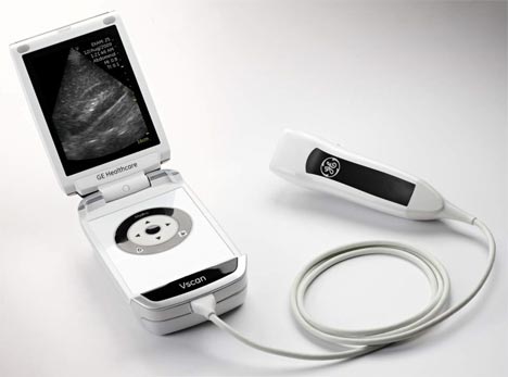
1-انتاج جهاز موجات فوق صوتيه بحجم الجوال
2-تطوير الأجهزة لتنتج صور ثلاثية الأبعاد
لا تستخدم الموجات فوق الصوتية للاغراض التشخيصية فقط ولكن تستخدم ايضا في الفحص الجموعي( الفحص الأولي) للامراض، وفي بعض الحالات تساعد كجزء من الوسيلة العلاجية.
اولا الفحص الجموعي:
تستخدم الموجات فوق الصوتية في الفحص الجموعي لامراض الاوعية الدمويةحيث تقيس مقدار تدفق الدم والانسداد في الشريان السباتي وبذلك تتوقع الاخطار الكامنة لحدوث السكتات الدماغية،ايضا تستخدم في قياس قطر الابهر البطني ولمعرفة وجود اي اتساع في قطره قد يؤدي لخطر تمزقه،وايضا يستخدم كفحص اولي للكشف عن حصوات المرارة او التهاباتها كما يستخدم في فحص الثدي للكشف عن وجود اورام سرطانية
.ثانيا الاستخدامات التشخيصية:
• اثناء الحمل : تستخدم مبدئيا في متابعة تقدم الحمل وتحديد وقت حدوثه كما يؤكد حيوية الجنين ومكانه طبيعيا داخل الرحم امخارجه وايضا يحدد مكان المشيمة بالنسبة لعنق الرحم ويحدد ايضا وجود عيوب خلقية أو تشوهات جسيمة من عدمه كما يحدد نمو الجنين وحركته ودقات قلبه وايضا يمكن من خلاله تحديد جنس الجنين بعد مرور 12 اسبوع من حدوث الحمل.
• امراض النساء: يتم بفحص الحوض لتشخيص اي اورام اوكبرفي حجم المبيض او الرحم او قناة فالوب ويتم الفحص اما بوضع المسبار علي جدار الحوض او بمسبار مخصص يوضع داخل المهبل.
• امراض القلب : يستخدم في تقييم وظائف عضلة القلب والصمامات ويبين وجود اي ضيق بداخلهم او ارتجاعوايضا يبين الدم المتدفق من خلالهم وايضا في تحديد كمية الدم التي يضخها القلب بالنسبة لكمية الدم الموجودة بداخله ويستخدم ايضا في تشخيص التهاب الشغاف او الغشاء المبطن لعضلة القلب من الداخل وايضا في تشخيص الرجفان الاذيني وذلك بما يسمي تصوير الصدى او الايكو وذلك اما بوضع مسبار الجهاز علي القفص الصدري او بداخل المرئ والطريقة الثانية تعطي صورة افضل لان القلب تشريحيا قريب جدا للمرئ .
• امراض الاوعية الدموية: يستخدم في الكشف عن وجود تجلطات دموية في الاوردة العميقة والسطحية كما يستخدم في تحديد ضيق الشرايين او اتساعها كما في الشريان السباتي لتشخيص حالات السكتة الدماغية
•:الامراض الباطنية :
يستخدم في فحص معظم اعضاء التجويف البطني مثل الكبد والطحال والبنكرياس كما يصور حصوات المرارة او اي انسداد في القنوات المرارية كما يستخدم في الكشف عن التهابات الزائدة الدودية او حصوات الكلي في المرضي الذين لا نستطيع تعريضهم لخطر الاشعة المقطعية مثل الحوامل والاطفال، ويعتبر افضل وسيلة تشخيصية في حالات التواء الخصية ويكشف عن اسباب تورم كيس الصفن اوآلام الخصيتين كما تستخدم الصور ثلاثية الأبعاد في الكشف المبكر عن الأورام الحميدة والخبيثة بالبروستاستا والقولون والمستقيم
• الامراض العضلية الهيكلية : يستخدم في الكشف عن تمزق الاربطة او امراض العضلات او اي نزيف داخل العضلات او المفاصل وأي اورام حميدة اوخبيثة كما يستخدم في الكشف عن التغيرات الروماتيزمية المبكرة.
• يستخدم في الكشف عن الغدة الدرقية ويحدد وجود اي اورام او عقد
ثالثا الاستخدامات العلاجية:
للموجات فوق الصوتية العديد من الاغراض العلاجية من اهمها:
تساعد في بعض العمليات مثل اخذ عينة من النسيج
وايضا تصريف السائل الصديدي الموجود بداخل الخراريج بواسطة الشفط بالإبر،وايضا يساعد في تحديد الاوردة الكبيرة في الرقبة وجدار القفص الصدري عندما يكون منالضروري تركيب تسريب وريدي في حين لا يمكن العثور علي وريد. .
استخدام الموجات الفوق صوتية المركزة ذات الشدة العالية في علاج بعض الاورام الحميدة
والخبيثة وهي ذات ترددات اقل من تلك المستخدمة في الاغراض التشخيصية.
• تستخدم في ايصال العلاج الكيمائي للخلايا السرطانية بالمخ والادوية الاخري للانسجة الاخري باستخدام الموجات ذات الترددات
العالية من 1- 10 ميجا هرتز ومجال من الشدة يتراوح بين 0-20 وات/سم مربع و طريقة عملها تعتمد علي تركيز هذه الموجات علي الخلايا لاثارتها لتجعلها تمرر الادوية أسرع.
• تستخدم مصدر موجات قوية تركز علي نسيج لتنتج تسخين موضعي به وتستخدم في العلاج الطبيعي وايضا في بعض علاجات الاورام السرطانية.
• تستخدم في المساعدة في الحصول علي البويضات من المبيض بتصوير المبيض اثناء ازالة البويضات للقيام بتلقيحها في ما يسمى باطفال الانابيب.
• تستخدم في علاج المياه البيضاء بتركيز الموجات علي العدسة لتذويبها.
• اكتشف مؤخرا قدرتها على تحفيز نمو العظام
وايضا في انفتاح الحائل الدموي الدماغي لتوصيل الدواء.
• تم اختبار الدوبلر في المساعدة في العلاج باستخدام منشط البلازمينوجين النسيجي في حالات السكتة الدماغية في عملية تسمي بتسريع إذابة الجلطات باستخدام الموجات الفوق صوتية.
اظهرت الموجات فوق الصوتية انها تعمل مع المضادات الحيوية تآزريا في قتل البكتيريا
• الخلايا حقيقية النواة المزروعة اكثر سماكة عن طريق الاسراع من اثبتت انها تجعل من امتصاص المغذيات.
كيف يتم تحضير المريض للقيام بفحص الموجات فوق الصوتية؟
التحضير لفحص الموجات فوق الصوتية بسيط عادة فمثلا للقيام بفحص المرارة يتجنب المريض الطعام فقط وليس شرب المياه لحوالي ست أو ثمان ساعات لتجنب انقباض المرارة نتيجة تناول الطعام مما يؤثر في النتائج، اما لفحص الحمل فيطلب من المرأة الحامل شرب من أربعة لستة اكواب من الماء علي الاقل قبل الفحص بساعة او ساعتين حتي تمتلئ المثانة .
الامعاء المليئة بالهواء تؤثر علي وضوح الرؤية، ويتم ازالة الملابس من الاماكن المطلوب عمل الموجات عليه حيث يوضع المسبار علي الجلد مباشرة بعد وضع الجل الخاص بها عليه لإبعاد الهواء.
مميزات وعيوب استخدام الموجات فوق الصوتية كعنصر تشخيصي
اولا المميزات:
• من اهم المميزات للموجات الفوق صوتية انها تكون صورة حية متحركة (REAL TIME) للنسيج او العضو المراد فحصه فمثلا تصور حركة القلب ومرور الدم فيه وتنقلها مباشرة على الشاشة.
الفوق صوتية عادة ما تكون غير جائرةعملية الفحص بالموجات
اي بدون حقن او إبر كما انها غير مؤلمة(NOT INVASIVE)
التصوير بالموجات فوق الصوتية متوفرة وسهلة الاستخدام
كما انها ارخص سعرا من وسائل التصوير الاخرى.
انها ليست إشعاع أيونيلاتسبب اي مشكلات صحية عكس الاشعة السينية والمقطعية حيث ويمكن ان تكرر عند الحاجة.•
تعتبر افضل من التصوير بالرنين المغناطيسي في حالات وجود منظم لضربات القلب عند المريض حيث انها لا تؤثر عليه، كما انها افضل منه في رؤية اوتارالعضلات.
ثانيا : العيوب:
غير مثبت انها ينتج عنها مخاطر طويلة المدى علي صحة المريض.
فقط ما يعتبر غير مستحب فيها إنها تحتاج الي تقني خبير حتى لايتم الحصول على نتائج غير دقيقة (FALSE RESULTS)
فمثلا يمكن الحصول على نتائج ايجابية عن وجود تشوهات بالجنين في حين انه لا يعاني من اي منها . | |
| | | | د.كمال سيد
Admin
عدد المساهمات : 2464
نقاط : 4252
السٌّمعَة : 9
الجنس :
علم بلدك :
تاريخ الميلاد : 03/04/1950
تاريخ التسجيل : 30/07/2012
العمر : 74
الموقع : السودان - سنار
العمل/الترفيه : طبيب عمومى وموجات صوتية
الساعة الان :
دعائي :
| | موضوع: رد: مقدمة تعريفية عن الموجات فوق الصوتية الثلاثاء أبريل 23, 2019 10:29 pm | |
| -What is sonography?
Ultrasonography, commonly called sonography, is a diagnostic medical procedure that uses high frequency sound waves (ultrasound) to produce dynamic visual images of organs, tissues, or blood flow inside the body. This type of procedure is often referred to as a sonogram or ultrasound scan. Sonography can be used to examine many parts of the body, such as the abdomen, breasts, female reproductive system, prostate, heart, and blood vessels.
Sonography is increasingly being used in the detection and treatment of heart disease, heart attack, and vascular disease that can lead to stroke. It is also used to guide fine needle, tissue biopsy to assist in taking a sample of cells from an organ for lab testing (for example, a test for cancer in breast tissue). Unlike X-rays, sonography is a radiation-free imaging modality.
The non-physician professionals who perform these procedures are known as sonographers and vascular technologists (who are sonographers specializing in imaging and tests of blood vessels).
There are several areas of specialization in the field of sonography.
These specialty areas are:
Abdomen - evaluation of all the soft tissues, blood vessels and organs of the abdominal cavities (for example, liver, spleen, urinary tract, pancreas)
Breast - frequently used to evaluate breast abnormalities that are found with screening or diagnostic mammography
Obstetrics/Gynecology - evaluation of the female reproductive system
Echocardiography - evaluation of the anatomy and hemodynamics (blood flow) of the heart, its valves and related blood vessels
Vascular Technology - evaluation and analysis of the hemodynamics (blood flow) of peripheral and abdominal blood vessels
Neurosonology - evaluation of the brain and spinal cord
Ophthalmology - evaluation of the eye, including orbital structures and muscles
What does a diagnostic medical sonographer do?
A diagnostic medical sonographer is a highly-skilled professional who uses specialized equipment to create images of structures inside the human body that are used by physicians to make a medical diagnosis. The process involves placing a small device called a transducer against the patient's skin near the body area to be imaged. The transducer works like a loudspeaker and microphone because it can transmit sound and receive sound.
The transducer sends a stream of high frequency sound waves into the body that bounce off the structures inside. The transducer detects sound waves as they bounce off the internal structures. Different structures in the body reflect these sound waves differently. These sounds are analyzed by a computer to make an image of the structure(s) on a television screen or that can be recorded on videotape.Sonographers have extensive, direct patient contact that may include performing some invasive procedures. They must be able to interact compassionately and effectively with people who range from healthy to critically ill. | |
| | | | د.كمال سيد
Admin
عدد المساهمات : 2464
نقاط : 4252
السٌّمعَة : 9
الجنس :
علم بلدك :
تاريخ الميلاد : 03/04/1950
تاريخ التسجيل : 30/07/2012
العمر : 74
الموقع : السودان - سنار
العمل/الترفيه : طبيب عمومى وموجات صوتية
الساعة الان :
دعائي :
| | موضوع: رد: مقدمة تعريفية عن الموجات فوق الصوتية الثلاثاء أبريل 23, 2019 10:30 pm | |
| table of contents
FAQS contd
Heart beat gone at 10 weeks.
Nuchal skin thickness.
White spots in the baby's bowel.
Shiny bright spot in the baby's heart.
Going back for a retake?
Slow heart rate of the fetus
Biochemical screening
A smaller abdominal circumference
Intracardiac echogenic foci
Enlarged kidneys
Left-handedness
3-D ultrasound
question
My wife has recently had a trans-vaginal scan and we were told that no heartbeat could be detected, she is 10 weeks pregnant by her dates. Her cycle varied between 28 & 32 days and we were told that the fetus was equivalent to 9 weeks only. We have been told to expect the worse for the repeat scan next week. How accurate is the scan relating to these matters and is it possible to miss the heartbeat at this stage? Will the fetus continue to grow without the heartbeat?
Answer:
Well, if the measurements indeed indicate a gestational age of 9 weeks, then it would be most unlikely not to be able to see the heart beat at this time. Very likely fetal demise has occurred. Don't expect the fetus will carry on growing without a pumping heart.
Question:
Can you tell me what the nuchal skin fold is for and what would be the use of measuring it during an ultrasound examination?
Answer:
The nuchal skin fold thickness or nuchal translucency is the thickness of the skin fold behind the nape of the neck. In chromosomal abnormalities this may be thickened due to venous or lymphatic engorgement. It is now almost routinely measured between 11 to 14 weeks to detect the presence of Down syndrome and fetuses with other chromosomal abnormalities.
Question:
I am 18 weeks at dates. I had an ultrasound scan last week and the technician told me that the baby's intestines looked whitier than they should be. She said she could not find any other peculiarity about my baby but suggested that I should come back for an amniocentesis this week. What does that mean? Does my baby have Down syndrome?
Answer:
I believe she is referring to 'echogenicities' in the fetal bowel which is considered to be one of the ultrasonic "soft signs" suggesting that the fetus may be suffering from chromosomal abnomalities the most common one of course is Down syndrome. The cause of the echogenicities is not clearly known but would appear to be due to calcified meconium or a maldevelopment of the gut of the fetus. The risk of Down syndrome in the presence of the findings of echogenic bowel is only 1.4% according to a study form the Benacerraf group in Boston. Other groups however had quoted a higher incidence. Counselling and genetic amniocentesis appear to be justified in the presence of such findings. A summary of this finding can be found here. Also check out the page Prenatal testing for Down Syndrome for related information.
Question:
I am going for a 'level 2' examination next week because my obstetrician said she could see a shiny bright spot in my fetus heart. I was told this could indicate Down syndrome. Is the risk really great? I am 15 weeks today.
Answer:
'Echogenicities' in the fetal heart is considered as another sonographic "soft sign" in the diagnosis of fetal chromosomal abnormalities. The echogenic areas probably represent calcifications at malformed areas in the papillary muscles of the heart. As reported by different authors, the presence of an intraventricular echogenic focus carries a relative risk of Down syndrome roughly 0 - 4 times as compared to a woman of similar age. Counselling, further ultrasonic assessment and amniocentesis will be considered in the presence of such findings. At the same time, the vast majority of fetuses who are chromosomally normal with an intraventricular echogenic focus do not have congenital heart disease.
Try to read this article in the Birth.com.au
Question:
I just had my 20 week ultrasound, and the technician told me everything looked fine, but he could not get a good look at the heart.?He wants to see me back in 3 weeks.?Is that routine?
Answer:
Yes. It is. Looking for abnormalities in the fetal heart is difficult and very much dependent on fetal position and clarity of the ultrasound picture at that time. It is common to ask patients to return for another scan particular the first examination was not reasurring of a normal cardiac configuration. Good sonologists or sonographers are very careful with malfornations. They won't let go until they are almost 100% certain things are normal or that the neccessary ultrasonic views have been taken.
Question:
I am 7 weeks pregnant. My scan yesterday showed that the fetal heart beat was only 80 per minute and the technologist said this was not good. She asked me to return for another scan next week. Should I be worred?
Answer:
A slow fetal heart beat is generally speaking significant and indicates unwell in the fetus. Normal heart rate at 6 weeks is 90-113 bpm and at 9 weeks is 144-170 bpm. At 5-8 weeks a bradycardia (<90 bpm) is associated with a high risk of spontaneous abortion of some 80%. (Benson CB, Doubilet PM. Radiology 1994; 192: 343-4). Other signs are also important in the assessment such as the size of the yolk sac and the amount of amniotic fluid.
ادخل الرابط
[rtl]http://hcp.obgyn.net/home[/rtl]
Question:
I know the question is not directly related to ultrasound but can you tell me a bit more about the 'double' and 'triple' test, and more recently the first trimester blood tests in the screening for Down syndrome?
Answer:
Read this informative article on biochemical screening for Down Syndrome.
Question:
I am currently 31 weeks pregnant. To date I have had 2 ultrasounds - one at 18 weeks, and one more recently at 30 weeks. According to the results of the latest the gestational age of the baby was 30 weeks, so my dates are correct. My concern is measurements of the baby - namely abdominal circumference. According to my doctor the head and femur measurements were above the 60th percentile, however the abdominal circumferance was only in the 9th. What concerns should I have regarding this? Is this considered abnormal intrauterine growth, and if so what may result? This is my second child. The first weighed 6lb 7oz at birth.
Answer:
This is a very common concern. Although in theory the baby appears to be asymmetrically small it can be interpretated as just a 'thin' baby. In general the birth weight of the second baby closely resembles the first. If your first baby was only 6 lb 7 oz then this baby is likely to have a similar weight. It doesn't sound 'thin' but figure-wise it is. The average weight of babies at term is around 7 lbs, so theoretically any weight below that will have an abdominal circumference falling below the 50th centile mark. 10th percentile at term is around 6 lbs so your results merely reflect that you will have a similar size baby as last time. Of course one should also pay attention to the amount of liquor and the exclusion of any congenital abormalitities.
Question:
I am 22 weeks pregnant and was told during my 20 week sonogram that my baby has a echogenic intracardiac focus in its left ventricle. Because of my timing, I was advised to have aminiocentesis for down syndrome. I am told my chances are slim because of my triple screen test was negative and my age of 29 years.
I would like to know what percentage and probability of my baby having the down syndrome.?I have been told this is still contraversial and it seems there is not much information out there. I am now waiting for my results from my aminio with great anxiety. Could you please advise me on this?
Answer:
Representing mineralisation of the papillary muscle, this soft sign for aneuploidy was not a common finding in this group of 6995 women (incidence 2.1%). The incidence of echogenic cardiac foci in 2nd trimester ultrasounds ranges from 0.17% to 7.4% in other studies. Estimates of the chance of Down Syndrome among fetuses with echogenic cardiac foci have varied from no increased risk up to a 4-fold increase in the age-related risk with several studies suggesting a risk of 1%. In one recent study of the 150 fetuses with the heart foci, 76% proceeded to karyotyping by chorionic villus sampling or amniocentisis or both and one had cordocentesis. The positive predictive value of an echogenic intracardiac focus for chromosomal abnormality in all patients was 3% (5/150). Only one of the five chromosomaly abnormal fetuses had an echogenic intracardiac focus as the only sonographic marker for chromosomal aberration.? Therefore, of the 92 cases where an echogenic focus was the only ultrasound marker, there was only one chromosomal abnormality (1%). Ninety-six percent of the echogenic foci were within the left ventricle.
With the controversy surrounding this ultrasound finding it is difficult to know what, if anything, is best fo each patients. Genetic Counseling is recommended. Many centers will not routinely recommend amniocentesis if this is an isolated finding and the patient is otherwise low risk.
You can also read this article on intraventricular echogenic focus.
Question:
I am 20 weeks pregnant and my doctor said that the baby had slightly enlarged kidneys. He was apparently referring to the inside measurements of the kidneys.?Could you tell me what this could mean? to my baby.
Answer:
The renal pelvis is the are in the kidney where urine collects before being passed down the ureter into the bladder. Renal pelvis dilatation, or pyelectasis is the term used to describe a visible renal pelvis, usually greater than 4 mm at 18 weeks. Most of them are transient and because they appear to disappear in time it is though that they are due to immaturity of the renal collecting system, and not harmful. Some are thought to arise from compression of the ureter as it descends into the pelvis and in to the pelvis. This may be a transient phenomenon but which may cause harm to the kidney over time.
Some of the dilatation appears to be due to reflux. In these cases, the urine flowing down into the bladder refluxes (comes back up the ureter) when the fetus empties its bladder. However renal anomalies or deviations from the normal renal anatomy have also been identified as markers for Down syndrome. It has been reported that the presence of isolated pyelectasis (enlarged collecting system) prenatally did not warrant amniocentesis for assessment of fetal karyotype. Subsequently, however, Nicolaides and associates reported that the presence of isolated pyelectasis was specifically associated with a 3-fold increase over the maternal age-related risk for chromosomal anomalies. In a 1996 evaluatio the association between isolated pyelectasis and an increased risk for Down syndrome was confirmed in gestational ages between 16 and 20 weeks, beginning at a maternal age of 31 years.
The incidence of chromosome abnormalities is greatest if any other structural malformation is present.
Most would agree that a renal pelvic diameter greater than 10 mm is more likely to have a worse outcome. Between 4 and 10 mm, it is more difficult to accurately predict the outcome. All fetuses with pyelectasis need a repeat scan later in pregnancy.
Question:
I have read from news magazines that there is an increase in left-handedness in babies in whom the mother had ultrasound scans a in pregnancy, and there may also be speech problems too. Is this true?
Answer:
Please refer to the Safety References for a discussion on the safety and possible harmful effects of Ultrasound..
[rtl]http://www.ob-ultrasound.net/joewoo3x.html[/rtl]
[rtl]http://www.ob-ultrasound.net/safenewn.htm[/rtl]
Question:
I was told that the 3-D scan will not give me more information as to whether my baby is normal or not than the usual 2-D ultrasound scan. Is this true and if that is the case why do we need the 3-D scan?
Answer:
3-D ultrasound has resulted largely from the advancement in computer technology and the vastly improving speed of the micro-processors. Indeed the benefits that 3-D has bought to ultrasound diagnosis has often been debated. The recognised advantages of 3-D over conventional 2-D ultrasound as it is at the turn of the millenium are its ability to enhance maternal-fetal bonding, improved comprehension of certain fetal anomalies by parents, Improved recognition and better confirmation of certain anomalies such as cleft lips, polydactyl, micrognathia, malformed ears, club foot, vertebral malformations and other anomalies appearing on the 'exterior' of the fetus, consequent to the benefit of volume and surface rendering. The development of transvaginal 3-D probes have further enhanced its value in the early diagnosis of congenital malformations.
In a recent article by Professor Asim Kurjak and his team in Europe, "Three-dimensional sonography in prenatal diagnosis: a luxury or a necessity?" (Journal of Perinatology, issue 3, 2000), he concluded,
".... the main advantages of three-dimensional ultrasound in perinatal medicine and antenatal diagnosis include scanning in the coronal plane, improved assessment of complex anatomic structures, surface analysis of minor defects, volumetric measuring of organs, "plastic" transparent imaging of fetal skeleton, spatial presentation of blood flow arborization and, finally, storage of scanned volumes and images. It is our decided opinion that three-dimensional sonography has gained a valuable place in prenatal diagnosis, becoming a necessity for every modern perinatal unit .... ".
Professor Stuart Campbell in London was one of the early proponents for the 3-D scan to be an important catalyst for mothers to bond to their babies. What are known as 're-assurance scans' and the perhaps misnamed entertainment scans' have started to develop. The attraction of being able to look at the face of your baby before birth was enthusiastically reported in lay parenting and health magazines. Manufacturers had adopted an unprecedented "profit marketing" strategy to advertise to providers and "reverse marketing" strategy to advertise to consumers, particularly after the arrival of the 4-D (dynamic or motion 3-D) machines.
Barbara Maier and her group (Horst Steiner, Alf Staudach etc.) in Austria reported in 1996 that mothers are more incentive to endure pregnancy-related difficulties, reduced anxiety, and improved capacity to cope. Dolores Pretorius in San Francisco reported in the same year that improved bonding between the mother and fetus could motivate mothers to refrain from smoking and other harmful behaviors during pregnancy. | |
| | | | د.كمال سيد
Admin
عدد المساهمات : 2464
نقاط : 4252
السٌّمعَة : 9
الجنس :
علم بلدك :
تاريخ الميلاد : 03/04/1950
تاريخ التسجيل : 30/07/2012
العمر : 74
الموقع : السودان - سنار
العمل/الترفيه : طبيب عمومى وموجات صوتية
الساعة الان :
دعائي :
| | موضوع: رد: مقدمة تعريفية عن الموجات فوق الصوتية الثلاثاء أبريل 23, 2019 10:30 pm | |
| frEQUENTLY ASKED QUESTIONS
table of contents
My bladder is bursting
Too early to see the fetus
Good to see the heart beating
Spotting in early pregnancy
Decreased fluid around the fetus
Polyhydramnios
Fetus is smaller than expected
Which date is more correct ?
Is my baby too small ?
I don't want to know the sex of my baby
Fail to recognize an ectopic pregnancy
Molar pregnancy
Too many scans
The sex of the baby
Heart rate and fetal gender
A cyst in the baby's brain
Just how accurate is ultrasound ?
Level II ultrasound
Routine ultrasound scan
Routine 13 weeks scan
FAQs
Question:
I am pregnant for 7 weeks. I went for a scan yesterday and was told I should have a full bladder. They made me drink 4 cups of water and my bladder was almost bursting at the end of the session. Is this really necessary ?
Answer:
A full bladder is usually necessary when the scan is done abdominally and in the early part of pregnancy. This would tend to lift the uterus up a little bit and being in front displaces the bowels away and act as an echo-lucent windows for the ultrasound to pass through. Ultrasound doesn't go through air (as in the bowels) well but transmits perfectly through liquid (as in the case of the urine in the bladder). Your doctor will be able to see things so much more clearly with the full bladder there especially the presence or absence of fetal heart beat. In a vaginal scan however, a full bladder will not be necessary.
Question:
I am pregnant for 6 weeks and 5 days, I turned up for a scan today but I was told they could not see the fetus. They say this could be due to my irregular menstruation, and ask me to return for another scan next week. What does that mean ? I am quite worried.
Answer:
Usually we should be able to see the fetal heart beat quite clearly by about 6 and a half weeks, particularly in a vaginal scan. By 6 1/2 weeks we mean 6 1/2 weeks from the last menstrual period (LMP), and not 6 1/2 weeks from the day of conception ( which is of course around the day of ovulation ). In a woman with irregular and slightly longish cycles (say, 35 days verses a usual 28 days), the day of ovulation would actually be postponed to 1 week on top of the 14 days in the usual case, that is, she ovulates on Day 21.
The number of days between the first day of menstruation and the date of ovulation (the follicular phase) is variable, but the number of days between ovulation and the start of the next menses (the luteal phase) is generally rather constant at about 14 days. So if you have a menstrual cycle of 42 days, then you ovulate on Day 28 ! Add 14 days and your next menses will come. Therefore if you have a longish cycle then you ovulate late and your fetus will be less advance then it should be, meaning it will appear smaller or cannot be seen at all.
Of course, if for other reasons, the fetus has not grown, then it will also show up smaller. The way to get around making a correct diagnosis of which is which is to repeat a scan in about a week's time, so that those who are smaller because of a date factor will then be found to have grown.
Question:
I am 7 weeks pregnant and had a scan yesterday. My doctor said everything is fine and he could see the heart beat. He said the chance of a miscarriage is very small and he looks quite confident when he said that. Is the comment valid ?
Answer:
Nowadays most Obstetricians would believe that the cause of the majority of 1st trimester miscarriages is a defect in the embryo, and is basically a chance event. Imagine a sperm coming together with an egg to develop into a fetus.This involves many millions of cell divisions and changes so that the chance of something going wrong is very great. If it happens very early on in development ( say 4 to 5 weeks ), the growth of the embryo may come to a stop.The heart will cease to beat. The body in due course rejects the non-viable material, giving rise to the process of 'miscarriage'. Therefore if by about 7 or 8 weeks, everything appears intact with a good going heart beat, it is quite reasonable to say that the pregnancy will most likely carry on. (The statistics is actually on an average better that 95%, although it will be lower in women with recurrent miscarriages).
Question:
My wife is 9 weeks pregnant with twins, she has on and off bleeding and spotting for the past 3 weeks. She already had 3 ultrasound scans, all of which showed two fetal heartbeats. we are concerned ( because of the spotting and bleeding ) which leads me to ask two 2 questions:
1. What are the known causes of spotting and bleeding?
2. Given a "positive" ultrasound, how does that weigh against the fact that there has been occasional spotting and bleeding?
Answer:
It is always difficult to pinpoint the exact cause of bleeding in a first trimester pregnancy. In your wife's case, it could just be bleeding from the cervix or the edge of the placenta ( which is bigger and more close to the cervix because of the twin pregnancy ) and these situations generally bear little or no significance. Sometimes the bleeding can carry on up to the second trimester. If the heartbeat is present, the same still holds i.e. over 97% of the time the pregnancy will carry on unharmed. In the old days any vaginal bleeding in the early part of pregnancy is called ' threatened abortion '. With the advent of Ultrasound, we have to say this is a misnomer, as in the cases when the fetal heartbeat is seen, the pregnancy is not ' threatened '.
عدل سابقا من قبل د.كمال سيد في الأحد مايو 12, 2024 10:30 am عدل 1 مرات | |
| | | | د.كمال سيد
Admin
عدد المساهمات : 2464
نقاط : 4252
السٌّمعَة : 9
الجنس :
علم بلدك :
تاريخ الميلاد : 03/04/1950
تاريخ التسجيل : 30/07/2012
العمر : 74
الموقع : السودان - سنار
العمل/الترفيه : طبيب عمومى وموجات صوتية
الساعة الان :
دعائي :
| | موضوع: رد: مقدمة تعريفية عن الموجات فوق الصوتية الثلاثاء أبريل 23, 2019 10:31 pm | |
| Question:
I had a scan yesterday. I am pregnant for about 30 weeks. My doctor said the fluid around the fetus is much decreased and told me to take more rest. He seems to look worried. Is this serious ?
Answer:
A decrease in the amount of fluid around the fetus (oligohydramnios) can be normal in most cases, and apparently can be improved with more rest, which in turn improves the circulation to the uterus.
A significant reduction in the amount of liquor, however, is associated with retardation of growth in the fetus and possible congenital malformations. Your doctor looked worried because he might be unsure as to whether the fetus is suffering from an abnormality or not. In such a case, referral to a centre with expertise in scanning for fetal malformations may be indicated. But remember, most cases would be quite normal and bed rest with improve things. The amount of liquor around the fetus does not have any relationship with the size of the mother or how much water she is drinking everyday.
Question:
My doctor has diagnosed polyhydramnios. I am currently 33 weeks gestation. I was sent to a specialist for a more advanced sonogram and told that nothing is wrong other than the polyhydramnios. I had a tri-screen at 18 weeks and it was negative as well. I still can't help but worry that something is wrong. My concern is that a birth defect undetected by a sonogram may still be a possibility and that the doctor may be keeping something from me. He is reluctant to do an amniocentesis. It seems that they want to give me as little information as possible and I can't help but wonder why if everything is o.k.
Answer:
A detailed ultrasound examination at 33 weeks should be able to diagnosis most, if not all the recognizable causes of polyhydramnios. A significant proportion of pregnancies with polyhydramnios does not have any demonstrable birth defects in the fetus. An amniocentesis may indeed be helpful for planning necessary treatment but your doctor probably felt that the chance of finding something wrong with the amnio is too small to warrant doing it. That is actually good news. I don't think your doctor is trying to keep things away from you.
Question:
My doctor said that my baby's head is a little 'smaller than dates' whereas the limb bone and the abdomen were quite on par with dates. Does that reflect that I have a baby with an abnormal head ?
Answer:
If this is your first scan, then it may be a problem with dates. If your dates have been quite well validated from a previous scan, then it appears your baby could have a slightly smaller head. But so what ? All of us has a different size head, isn't it ? And mind you, your doctor measures the side-to-side 'diameter' of the fetal head and if your fetus has a relatively 'flatter' head it will appear 'smaller' on measurement. This may simply reflect the head's configuration rather than anything that you should be alarmed about.
It will need a size lag of up to 3-4 weeks before one would start to consider malformations such as microcephaly ( small head syndrome ).
Question:
I had a scan at 8 weeks and my doctor said the fetus is only 7 weeks, and it was because I had irregular menstrual cycles. I had another scan at 34 weeks and my doctor now said the fetus size was only 31 weeks. Should I stick to the earlier scan or should I use the later scan to fix my Due date ? If my fetus is smaller, would it be wise to leave it in the womb for as long as possible so that it can grow bigger and get more mature ?
Answer:
You should always use the result of a scan that is done earlier on in pregnancy for 'dating' purposes as it will be more accurate. In the later part of pregnancy the measurements will be affected by growth variations and will no longer reflect the fetal 'age' correctly. In your case, the findings apparently indicated that you have a smaller than average baby. It is incorrect logic to think that babies who are not growing well should be left inside the womb for a longer period of time. In fact the reverse should be true. The baby may be better off outside than in and for this reason we sometimes have to deliver these babies well before the actual due date. | |
| | | | د.كمال سيد
Admin
عدد المساهمات : 2464
نقاط : 4252
السٌّمعَة : 9
الجنس :
علم بلدك :
تاريخ الميلاد : 03/04/1950
تاريخ التسجيل : 30/07/2012
العمر : 74
الموقع : السودان - سنار
العمل/الترفيه : طبيب عمومى وموجات صوتية
الساعة الان :
دعائي :
| | موضوع: رد: مقدمة تعريفية عن الموجات فوق الصوتية الثلاثاء أبريل 23, 2019 10:32 pm | |
| Question:
I had a scan at 34 weeks. My doctor said the estimated weight of the fetus is 2.0 Kg. Is this too small? How much should an average baby weigh at 34 weeks ?
Answer:
The weight for an average size fetus at 34 weeks is about 2.2 Kg. 2.0 Kg will be on the 25th percentile line. We would say a baby is small-for-dates when it's weight is below the 10th percentile line, and in the case of 34 weeks this will be 1.8 Kg. Please take a look at the Intrauterine weight chart. Your baby is on the small side, but not too small. You must also know that estimating the weight of the fetus with ultrasound measurements sometimes can incur errors of over 10 percent. Your doctor will usually make an assessment together with other ultrasound findings.
Question:
I will have a scan tomorrow. What is the chance that I would be able to see the sex of my baby on the screen? I really do not want to know this before the baby is born.
Answer:
The chance that you will recognize the genital organs of the baby without any prompting is very small. Interpretating an ultrasound image requires a lot of training and skill and is not like looking at a photograph of a person taken in front of him. Patients are often unable to 'see' the parts of the fetus the doctor is actually carefully studying during a scanning session.
Question:
A friend of mine was seen at the ER because of pain. She was pregnant for about 7 weeks. They did a scan and found no sac in the uterus and asked her to go home. Two days later she was admitted again to another hospital with massive bleeding from an ectopic pregnancy. Why was this initially missed on the scan ?
Answer:
Ectopic pregnancyis notorious for not being diagnosed early, as the physician has always to exclude basically 3 other conditions in early pregnancy which give rise to pain.
1. She can have pain for no apparent pathology in early pregnancy, probably as a result of of uterine and venous engorgement.
2. Missed or inevitable abortion.
3. Ruptured ovarian cyst or pain arising from other abdominal organs.
Ultrasound, unfortunately, will not be able to make a 'definitive' diagnosis of ectopic pregnancy in most of the cases as it is often not possible to visualize the actual gestational sac in the fallopian tubes. The fact that there is no gestational sac seen in the uterine cavity may just mean that the pregnancy has not been growing normally or that some sort of an abortion might have occured or that the pregnancy, due to to an irregular menstrual cycle in the woman, is much less advance than is thought.
The doctor often needs to take into account of the menstrual history, the time she had a positive pregnancy test, the intensity of the pain, physical signs, a blood count and findings on ultrasound scan.
Question:
I would appreciate any information on molar pregnancy that you can provide. Last year I had emergancy surgery done because of this condition. I was told very little information about it at the time. I was wondering specifically: what exactly does it mean to have a molar pregnancy, what causes this, what are the chances of having one, and is normal conception/pregnancy difficult for someone who has had one. Again, any information on previous studies of this condition is appreciated.
Answer:
I am afraid I won't be able to give you detailed descriptions about a specific disease. May I suggest that you use the search engine "Google" or "Alta Vista" to find the answers you need.
[rtl]http://www.google.com/[/rtl]
[rtl]http://www.altavista.com/[/rtl]
You can also use the DejaNews engine to search the Newsgroups.
[rtl]http://www.DejaNews.com/[/rtl]
I am sure you will find plenty of information that you need.
Question:
My wife has already had 5 scans in her present pregnancy because our doctor thinks the baby is not growing very well. I am worried if this may be harmful to the baby. Can you tell me where I can find some information concerning the safety of ultrasound in pregnancy? Also, why does she need to be scanned so many times ?
Answer:
Please read the section on Safety references. There is presently no evidence of a cumulative effect of ultrasound in the fetus. Your doctor probably wanted to monitor the growth of the fetus by taking body measurements at set intervals. If the growth lag is getting serious, he would have to perform some other tests to monitor the wellbeing of the fetus and perhaps act promptly to deliver the baby at an earlier date. | |
| | | | د.كمال سيد
Admin
عدد المساهمات : 2464
نقاط : 4252
السٌّمعَة : 9
الجنس :
علم بلدك :
تاريخ الميلاد : 03/04/1950
تاريخ التسجيل : 30/07/2012
العمر : 74
الموقع : السودان - سنار
العمل/الترفيه : طبيب عمومى وموجات صوتية
الساعة الان :
دعائي :
| | موضوع: رد: مقدمة تعريفية عن الموجات فوق الصوتية الثلاثاء أبريل 23, 2019 10:32 pm | |
| Question:
I went for my ultrasound yesterday (20 weeks and 2 days). I also had one at 17 weeks and the technician said she did not see any boy parts, and advised me to come back for a repeat scan. Well I went yesterday, and she said there were definitely no boy parts down there, and she started looking for the labia. She could'nt see the labia after searching for about 10 minutes and told me that since by this time they can almost always see the penis or scrotum, she felt this baby is a girl, although she didn't seem to look that reassuring. We were able to tell our sons were boys at 18 and 20 weeks so I am wondering why this time it's so difficult. Is it unusual not to see the labia at 20 weeks, and does that mean it can still be a boy ?
Answer:
I can see that you are quite anxious to know the sex of your baby. In order to be certain the sonographer should reassure himself that he actually sees the penis and scrotum in the case of a boy and the labia in the case of a girl. The absence of the penis must not be taken as sufficient evidence of the fetus being a girl. Your sonographer was careful not to make a mistake which could be extremely embarrassing. Most of the time one should be able to tell the sex of the baby by about 20 weeks and very often even at sixteen. On the other hand, as the correct visualization of any fetal part depends of a host of factors such as fetal position, amount of liquor and thickness of the abdominal wall, I have had occasions when I cannot be certain about the sex even at 28 weeks.
I have been repeated asked many many times about just how accurate is the diagnosis of fetal gender by ultrasound? Well, I have to say that it should be very accurate (somewhere between 95 and a hundred percent), particularly after about 20 weeks, or even sixteen. However I must say again that one must remember the sonologist may not always get a good view of the perineum (private parts) of the fetus for a variety of reasons and therefore may not furnish you with an "accurate" answer. One really needs to put this question to the person who performs the scan. He or she is the only person to be in the position to tell you just how accurate "that" particular diagnosis is.
Question:
I have heard that the heart rate of the fetus is slower when the fetus is male ( less than 140 per minute ), is this true and when is the best time to count the heart beat to tell the difference ?
Answer:
There is no scientific basis for a difference in heart rate between fetuses of the two sexes. It is another of maternity's many myths. Please read a very good article on this subject by Terry DuBose.
Question:
I am 18 weeks pregnant. I went for a routine scan yesterday and I was told my baby has a small cyst in the brain. My doctor said she would like to scan me again in 2 weeks time to see if the cyst grows any bigger and if there are other things wrong with my baby. She mentioned something about chromosomes. Is my baby going to be very sick or mentally retarded ?
Answer:
I think your doctor probably saw a "choroid plexus cyst" in the fetal head. This occurs in about 0.5 to 1 percent of fetuses and the great majority of them will disappear eventually. They have, however been found to be associated with chromosomal abnormalities notably Trisomy 18 and trisomy 21 (some 5 to 30 percent of these fetuses have the cysts). It is probably worse if there are more than one cyst and they are bigger than 10mm. Single cyst is almost always benign. It is important to diligently check for other features of chromosomal trisomies in the subsequent scan. An amniocentesis may be ordered if findings are in doubt.
Try to read this article in the Birth.com.au
Question:
Can you tell me just how accurate is ultrasound ? I heard that abnormalities can sometimes be missed by a scan.
Answer:
There is no definite estimate of exactly how accurate ultrasound is at detecting disabilities, and there are both false negatives and false positives.
As far as detecting fetal abnormalities goes, several points have to be bornt in mind :
a) Not every problem can be diagnosed with ultrasound -- conditions which do not manifest as a structural or gross functional abnormality (such as a very abnormal heart beat or mental retardation) may be missed.
b) Not every problem which can be detected will be diagnosed. For example, in many ultrasound examinations the baby's fingers and toes are not counted, even though it is possible to do so should it be necessary.
c) The ability to detect abnormality in the fetus at an ultrasound exam depends on a number of factors, such as the size and position of the fetus, the amount of amniotic fluid around the fetus, the body habitus of the mother (whether she is fat or slim), the type of equipment used, and, most importantly, the skill and experience of the operator.
d) Some problems (such as anencephaly) are more readily diagnosed than others (such as cleft palate).
The accuracy of ultrasound for dating a pregnancy depends on at what point during the pregnancy the ultrasound is taken. Pregnancy dating is most accurate during the first eighteen weeks of pregnancy. Measurement of the sac at five to seven weeks is not accurate. Measuring the crown-rump length gives an accuracy of plus or minus three days at seven weeks; this test can be used from the seventh to the fourteenth week. Between fourteen and twenty-six weeks, measurements of the biparietal diameter, the femur length and abdominal circumference are generally used. The accuracy is plus or minus seven to ten days. The accuracy further declines with advancing gestation age. | |
| | | | د.كمال سيد
Admin
عدد المساهمات : 2464
نقاط : 4252
السٌّمعَة : 9
الجنس :
علم بلدك :
تاريخ الميلاد : 03/04/1950
تاريخ التسجيل : 30/07/2012
العمر : 74
الموقع : السودان - سنار
العمل/الترفيه : طبيب عمومى وموجات صوتية
الساعة الان :
دعائي :
| | موضوع: رد: مقدمة تعريفية عن الموجات فوق الصوتية الثلاثاء أبريل 23, 2019 10:33 pm | |
| Question:
What is meant by a Level II ultrasound examination?
Answer:
A level II ultrasound examination refers to a 'targeted' examination. This is usually performed at a perinatal center where more expertise in ultrasound scanning is available. Situations when a level II scan is ordered include:
suspected fetal anomaly during a level I examination, severe IUGR, elevated maternal-serum AFP, Oligohydramnios, polyhydramnios, two-vessel umbilical cord detected at the level I examination, fetal cardiac arrhythmia, and exposure to drugs or chemicals in the first trimester....etc. Detailed scanning for any congenital anomalies and growth abnormalities is done on top of the basic documentations obtained at the level I examination, which refers to:
in the first trimester,
a. documentation of the location of the gestational sac.
b. documentation of the crown-rump length.
c. documentation of the presence or absence of fetal life.
d. evaluation of the uterus (including cervix) and adnexa.
in the second and third trimester,
a. documentation of fetal life, number, and presentation.
b. documentation of an estimate of amniotic fluid volume.
c. documentation of placental location.
d. documentation of gestational age. Assessment of the BPD, FL, AC and othe fetal parameters.
e. documentation of basic fetal anatomy (including a basic 4 chamber view of the heart, spine, stomach, kidneys, bladder, umbilical cord insertion ... etc ).
One should not not dwell too much on the definitions or guildlines for a level II ultrasound scan. In a level II scan the sonologist should try as hard as he can to look for and assess any abnormality that is present in the fetus. It's meaningless to be thinking about level III or even level IV scans.
Question:
My wife never had a scan for her present pregnancy. I have heard that in many hospitals an ultrasound scan at about 20 weeks is routine. Why is it not done in my wife's case ?
Answer:
The issue of 'Routine scans' in a 'low risk' patient is still a matter of some debate at the moment. I personally is for the implementation of the policy of a routine scan for every pregnancy at 18-20 weeks. Few people will doubt it's usefulness in improving gestational age dating, in the early recognition of fetal anomalies, in the identification of intrauterine growth retardation and of multiple gestations.
There is apparently a distinct difference in the attitude towards routine ultrasound screening between the Europeans and the Americans. Routine screening scans were introduced in Germany in 1980, in Norway in 1986 and in Icelend in 1987. The scans basically try to date the pregnancy, exclude twins and detect any fetal malformations that may be present. In the U.S., routine scans in pregnancy has however been looked upon with much controversy and their cost-effectiveness and validity in improving 'quantifiable' perinatal outcome has not been firmly established. Nevertheless, those who argue for a routine scan would claim that parents have a natural desire to know if any fetal congenital anomaly is present or if there is any health compromise in the fetus. Being able to reassure the parents is a natural part of prenatal care.
Two important large scale studies emerged to address the issue: the RADIUS study (Routine Antenatal Diagnostic Imaging with Ultrasound) with a cohort of 15,000 low-risk pregnancies in the United States in 1993 and the Eurofetus Study in Europe in 1997 in which 200,000 low-risk pregnant women in 60 hospitals had obstetric ultrasound examinations performed in centers proficient in prenatal diagnosis. A 61% overall detection rate of structural anomalies in the Eurofetus study contrasted sharply with the 35% overall detection rate in the RADIUS study.
In order to address the conflicting data and conflicting opinions on this topic, a conference was held at The Rockefeller University in New York City, in June 1997, sponsored by the New York Academy of Sciences. Over 150 scientists and clinicians participated in the meeting, with highly informative presentations and discussions.
Apparently the following conclusion was made:
" .... In summary .... we have tried to put together comprehensive state-of-the-art information on the routine obstetric ultrasound controversy. Our conclusion .... is that routine obstetric ultrasound is warranted for all pregnancies, but only if it is performed in a quality manner. Although there is still scientific and economic controversy about our conclusion, we would argue that, at a minimum, there is an ethical obligation to present the option of an 18-22-week routine obstetric ultrasound examination in clinical centers in which quality ultrasound is available. We hope that our efforts will move public policy in this direction and encourage further discourse on this most important topic in contemporary obstetrics."
In a setting with routine scanning, the cost of healthcare could be greatly increased. Such cost-effectiveness consideration is likely to be of concern at the hospital where your wife is attending. For an individual private patient I have absolutely little hesitation of ordering a scan at 18-20 weeks on a routine basis.
It should also be bornt in mind that over half of the significant abnormalities in the fetus could be missed on a routine scan. This will generate a false sense of assurance to the parents. Nevertheless, this does not mean the situation would be any better without this scan.
Please also read another of my discussions Here, and
the RCOG statements on routine ultrasound scans in pregnancy.
Question:
Is there any advantage of doing an earlier screening scan in pregnancy (12 weeks) to detect anomalies, or is it preferable to wait for the regular scan at 18-20 week.?
Answer:
It is noe becoming almost routine to perform a scan at around 11-14 weeks, mainly to look for an increased nuchal translucency and absent fetal nasal bone for the detection of chromosomal anomalies. With the documentation of an absent nasal bone and a thickened nuchal translucency in the fetus the chance of Trisomy 21 is much increased. More recently the presence of triscuspid regurgitation are also looked at to increase the detection rate.
The screening is usually performed with the abdominal rather than the vaginal scan. A repeat scan at about 18 to 20 weeks will still be necessary for such anomalies as cardiac, spinal, intracranial and facial abnormalities.
[rtl]http://www.ob-ultrasound.net/joewoo3.html#1a[/rtl] | |
| | | | | | مقدمة تعريفية عن الموجات فوق الصوتية | |
|
مواضيع مماثلة | |
|
| | صلاحيات هذا المنتدى: | لاتستطيع الرد على المواضيع في هذا المنتدى
| |
| |
| |
|