عدد المساهمات : 2464نقاط : 4252السٌّمعَة : 9الجنس : علم بلدك : تاريخ الميلاد : 03/04/1950تاريخ التسجيل : 30/07/2012العمر : 74 الموقع : السودان - سنارالعمل/الترفيه : طبيب عمومى وموجات صوتيةالساعة الان : دعائي :
موضوع: Doppler US الثلاثاء أبريل 30, 2024 4:33 pm
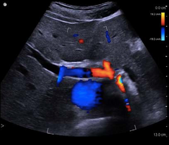
Doppler ultrasound is a noninvasive test that can be used to measure the blood flow through your blood vessels. It works by bouncing high-frequency sound waves off red blood cells that are circulating in the bloodstream. A regular ultrasound uses sound waves to produce images, but can't show blood flow. Doppler ultrasonography is medical ultrasonography that employs the Doppler effect to perform imaging of the movement of tissues and body fluids, and their relative velocity to the probe. Wikipedia The Doppler effect (also Doppler shift) is the change in the frequency of a wave in relation to an observer who is moving relative to the source of the wave. The Doppler effect is the change in frequency that you hear when a source and an observer are moving with respect to each other. The classic case is when you have an ambulance racing by you: the pitch of the siren is higher when it is moving toward you and lower as it goes away.ما هو التخطيط فوق الصوتي (دوبلر)؟ التصوير فوق الصوتي بالدوبلر اختبار غير متوغل (بدون جراحة) يُمكن استخدامه لقياس تدفق الدم في الأوعية الدموية. ويعمل عن طريق ارتداد الموجات الصوتية عاليَة التردد من خلايا الدم الحمراء التي تسري في مجرى الدم. ويستخدم جهاز أشعة دوبلر للحامل لسماع صوت نبضات قلب الجنين مثل أي جهاز يستخدم أشعة الموجات فوق الصوتية، حيث يقوم الطبيب بعمل الفحص الروتيني في بداية فترة الحمل (قبل مضي 12 أسبوعا من الحمل) للتأكد من صحة الجنين ومن أن ضربات القلب لديه طبيعية، وعادةً ما يتم إجراء اختبار أشعة دوبلر للحامل خلال الثلث الثاني من الحمل يُستخدم دوبلر الجنين للاستماع إلى نبضات قلبه بدايةً من أواخر الأشهر الثلاثة الأولى من الحمل كإجراء روتيني حتى موعد الولادة. يستطيع دوبلر تقييم حالة الدورة الدموية في الجنين والرحم والمشيمة. لذلك نلجأ إلى استخدامه بصفة أوسع مع حالات الحمل الحرجة وعالية الخطورة عند القلق بشأن حالة الطفل. (Doppler Ultrasound, Dupplex) هناك أيضًا أجهزة دوبلر صغيرة تهدف لفحص تدفق الدم، ويستخدم مصطلح دوبلكس (Dupplex) لوصف جهاز دوبلر الذي يكوّن صورة ثنائية الأبعاد على عكس دوبلر العادي الذي يوفر معلومات ذات بعد واحد ما هو التصوير بالموجات فوق الصوتية ؟ التصوير بالموجات فوق الصوتية هو فحص الجسم fموجات صوتية عالية التردد ثم التقاط الموجات المرتدة من الأنسجة وتحويلها إلى صورة مرئية. يمكن إستخدام هذه التقنية لإظهار بنية وحركة أعضاء الجسم الداخلية، فضلا عن تدفق الدم من خلال الأوعية الدموية. هل فحص الدوبلر الملون ضروري للحامل؟ ضعف في وصول التغذية والأكسجين إلى الجنين بشكل جيد بفضل هذا الاختبار سيكون طبيبك قادرًا على الاكتشاف المبكر في حالة وجود أمراض القلب ,تسمم الحمل , انسداد الاوعيه الدمويه او التشوهات فحص تشوهات الجنين في الثلث الاول من الحمل: ويسمى الفحص المشترك (Combined First Trimester) حيث يتم عمله بين الاسبوع 11 والاسبوع 14 من الحمل وهو الفحص المعتمد حالياَ عالمياَ بسبب ارتفاع دقته من حيث الكشف عن التشوهات الخلقية التي تصل الى حوالي 90%-93% حسب الدراسات،
عدل سابقا من قبل د.كمال سيد في الأربعاء مايو 01, 2024 8:45 am عدل 4 مرات
د.كمال سيد Admin
عدد المساهمات : 2464نقاط : 4252السٌّمعَة : 9الجنس : علم بلدك : تاريخ الميلاد : 03/04/1950تاريخ التسجيل : 30/07/2012العمر : 74 الموقع : السودان - سنارالعمل/الترفيه : طبيب عمومى وموجات صوتيةالساعة الان : دعائي :
موضوع: Doppler Effect الثلاثاء أبريل 30, 2024 8:35 pm
The Doppler effect is an essential concept for studying sound waves and frequencies. It is applicable in several scientific disciplines such as aeronautics, planetary science, and similar subjects. Christian Johann Doppler first introduced it in 1842. Before understanding the application of the Doppler Effect, let us first go through the fundamentals of the Doppler Effect.
The Doppler Effect Basics
Also known as the doppler shift, the doppler effect results from a change in the frequency of a wave from the perspective (point of view) of an observer moving at a speedrelative to the source of the wave. A common example of the Doppler effect is the shift in the pitch of a vehicle’s horn sound as it moves forward in the same direction. The Doppler effect takes place because 1/ each successive sound wave coming from the position 2/ gets closer to the observer. Therefore, 3/ it takes less time for each wave to move towards the observer than the previous wave.
Applications of the Doppler Effect
Let us now look at the following points to understand the application of the Doppler effect in various fields –
Vibration measurement
The doppler effect is also used in measuring vibration through the laser Doppler vibrometer. It is equipment that helps measure vibration by generating a laser beam moving towards the object’s surface.
Audio equipment
The Doppler Effect is used in some speakers that run on an electric motor to generate acoustics surrounding a loudspeaker. Due to this generation of acoustics, the sound generated from speakers moves in a circle resulting in fast-changing frequencies.
Radar
The most common application of the doppler effect includes radars. A few radars use the doppler effect to determine the velocity of the target object. Investigation departments and police frequently used the radar to identify vehicles running at high speed and similar actions requiring the usage of a radar.
Diagnosis and Treatment
The Doppler Effect also finds its application in the medical field in identifying vascular problems, including stenosis. The main reason for the application is the limitations of the echocardiogram, which is used to generate an accurate evaluation of the velocity of cardiac tissues and blood. It identifies the direction in blood flows at a given point in time.
Military
The military uses the popular effect to identify a submarine’s speed and other equipment with the help of Sonars. The buoy (floating object) is responsible for generating consistent and stable frequencies producing the Doppler Effect every time they come near an object in motion near the submarines. It also helps to record the velocity resulting from the object in motion every time it approaches the target object.
Aerospace Navigation
Since radars are used in aerospace navigation, Doppler Effect helps calculate the speed of flying equipment, including drones, plane jets, and any other object frequently used for aerospace missions. Therefore, it can be said that The Doppler Effect is also applicable in aeronautical missions for the calculation of objects’ speed.
Astronomy
Astronomy is another significant application of the doppler effect to calculate the speed at which stars rotate and if they are in a close range. This application is required because telescopes used for astronomical purposes have their limitations. They may not be able to serve the purpose when the given distance images are not appropriately clear.
عدد المساهمات : 2464نقاط : 4252السٌّمعَة : 9الجنس : علم بلدك : تاريخ الميلاد : 03/04/1950تاريخ التسجيل : 30/07/2012العمر : 74 الموقع : السودان - سنارالعمل/الترفيه : طبيب عمومى وموجات صوتيةالساعة الان : دعائي :
موضوع: Doppler Effect الثلاثاء أبريل 30, 2024 8:55 pm
The Doppler Effect and Sound Waves
This phenomenon explains the concept of the doppler effect in sound concerning the response of shockwaves. These are produced due to the movement of source waves and an upward shift in the frequency when the source and observer are receding. In this case, the Doppler effect is noticed when the speed of the primary source is relatively slower than the speed of the sound waves. However, if the primary source starts to move along at the same speed, the process is no longer the same as before.
The Relativistic Doppler Effect
The relativistic effect explains the doppler effect with light concerning a fluctuation in the frequency of light due to the relative motion of the source and the observer. It is quite the opposite of the non-relativistic Doppler effect, and the equations usually include the time dilation effect of special relativity. It happens when the source moves towards the observer and perceived frequency goes above the levels of emitted frequency.
Conclusion
This article explains the application of the Doppler effect. Also known as the Doppler Shift, the Doppler Effect refers to the change in frequency of a moving wave concerning the relative motion of the observer and the wave source.. The Doppler Effect is applicable in flow measurement, vibration measurement, velocity profile measurement, radar, and many more use cases.
The Doppler effect occurs because each wave pattern generated from a position relatively closer to the observer starts moving towards the source of the waves. Due to this phenomenon, it takes slightly less time for each wave to reach the observer.
The Doppler effect applies to light because it also affects the light coming from other objects located nearby. It is usually applicable in light waves where a body in the space is considered blue-shifted if the light waves are close. On the other hand, it is considered red-shifted if the light waves are spread widely and drifting away.
عدد المساهمات : 2464نقاط : 4252السٌّمعَة : 9الجنس : علم بلدك : تاريخ الميلاد : 03/04/1950تاريخ التسجيل : 30/07/2012العمر : 74 الموقع : السودان - سنارالعمل/الترفيه : طبيب عمومى وموجات صوتيةالساعة الان : دعائي :
موضوع: Doppler Applications الأربعاء مايو 01, 2024 10:37 am
How It Works: Doppler Ultrasound Imaging What is Doppler Imaging? Ultrasound doppler imaging is the ability to estimate and measure blood flow through various veins, arteries and vessels. Generally portrayed as a moving picture on an ultrasound system screen, one can usually recognize a doppler test from the color flow that is visible on the ultrasound image. The color in the image can be interpreted based on measuring blood movement in the specific area being photographed. Doppler imaging is different than conventional ultrasound imaging in one fundamental way: It doesn’t actually image any structures. Conventional ultrasounds provide images for various structures, organs and veins to diagnose growths, breaks, structural problems, and many other potential ailments. Doppler imaging, on the other hand, projects images simply of blood flow.
Due to the non-invasive and non- radioactive nature of the ultrasound doppler imaging, it is a methodology that is globally established and revered (respected). Instead of utilizing radiation or invasive features, the doppler functions the same way as other ultrasound imaging equipment does; employing high pitched sound waves that reflect and are translated into colors, images and various movements . Doppler Imaging’s Service: Doppler imaging is different than conventional ultrasound imaging in one fundamental way: It doesn’t actually image any structures. Conventional ultrasounds provide images for various structures, organs and veins to diagnose growths, breaks, structural problems, and many other potential ailments. Doppler imaging, on the other hand, is applied to detect blood flow and various potential hazards that can occur within the veins, arteries and vessels. Doppler imaging is generally used to detect blood clots, identify poorly functioning valves in veins, determine whether an artery is blocked, or recognize decreased blood circulation throughout the body. All of these potential threats to one’s health and life can be observed and prevented through use of doppler imaging There are different applications that people employ doppler imaging for: cardiac doppler, for example, and examining the blood flow to and from the heart is popular and extremely crucial part of cardiological examinations.
عدد المساهمات : 2464نقاط : 4252السٌّمعَة : 9الجنس : علم بلدك : تاريخ الميلاد : 03/04/1950تاريخ التسجيل : 30/07/2012العمر : 74 الموقع : السودان - سنارالعمل/الترفيه : طبيب عمومى وموجات صوتيةالساعة الان : دعائي :
موضوع: Doppler modes الأربعاء مايو 01, 2024 3:36 pm
Understanding Different Doppler Imaging Modes: Once upon a time, before modern ultrasound technology, it was considered advanced for an ultrasound system to have color doppler imaging. Color doppler has two colors, red and blue. Using these colors, it became easier to perform exams and diagnose ailments accurately. Some older ultrasound systems, however, don’t have color doppler, and rely on a black and white screen to diagnose patients. Nowadays, thankfully, nearly all ultrasound machines uses color doppler. In order to understand how doppler imaging works, one must understand how to identify what the colors on the ultrasound system indicate. The 1/ colors on the screen represent blood flow that is in motion. 2/ Blood moving towards the transducer is red, and blood moving away is blue. 3/ The lighter the shade of red or blue, the more rapid and quick the blood flow. 4/ The darker the shade, however, the slower the blood is moving. Power doppler, a newer and more advanced form of doppler imaging, is used to provide doppler images for areas that are more difficult for conventional doppler modes have a difficult time capturing. It is used for small vessels, kidneys, and the brain. Although it has become more common in newer ultrasound systems, not all ultrasound machines employ power doppler options Doppler Transducers: Every ultrasound manufacturer makes their own doppler probes, generally named “CW” (standing for continuous wave, a description of doppler images), followed by the frequency range. For example, the Mindray CW2s – which is short for Mindray continuous wave 2 MHz. probe. Most doppler probes, regardless of the manufacturer, are given names that are all similar to this example. Doppler transducers have several other nicknames that individuals in the medical world refer to them as. Some of these nicknames include “blind probes”, “pedoff probes”, “continuous wave probes”, and obviously “doppler probes”. Any of these names are common among the medical community, and are used interchangeably. Many states and insurance companies within the continental United States require that a diagnostician or sonographer that is purchasing an ultrasound machine must include a doppler continuous wave transducer with the machine. This requirement further emphasizes the importance of employing doppler imaging.
Ultrasound Safety: There is always the concern, particularly when it comes to medical equipment, regarding the safety of the devices and the potential side effects that occur as a result of employing these machines. It is important to know that ultrasound machines are known to be completely harmless. They function through the utilization of sound waves emitting from the probe or transducer. These sound waves are then translated into images by the ultrasound machine or system. There is no need to fear when using ultrasound machines – it is a quick, easy and efficient way to diagnose and help determine a patient’s ails.
عدد المساهمات : 2464نقاط : 4252السٌّمعَة : 9الجنس : علم بلدك : تاريخ الميلاد : 03/04/1950تاريخ التسجيل : 30/07/2012العمر : 74 الموقع : السودان - سنارالعمل/الترفيه : طبيب عمومى وموجات صوتيةالساعة الان : دعائي :
موضوع: Basic US Physics الأربعاء مايو 01, 2024 9:15 pm
General principles of US Physics
This is a general topic in which is discussed Basic US physics, important definitions, the differnt US modes, the normal sonographic anatomy & patology, IMPORTANT DEFINITIONS 1/GREY SCALE Is a display mode in which echo intensity is recorded as degrees of brightness or shades of grey US image formation depends on assesment of the echo strength & Go (pulse) Return (echo) time if returning echo is very strong it appears very bright (hyperechoic), & if weak it appears grey (hypoecoic), & if no echo it appears black (Anechoic) in the screen. SO this defines grey scale 2/Echogenic any area of US which produces echo is called echogenic. 3/Anechoic or echofree Areas without echoes are called Anechoic or echofree i.e no US echo returns back to TXR. All cavities containing fluid produce NO echoes e.g UB (urine) & GB (bile). A simple cyst is Anechoic = echofree (NO echoes returning back to TXR) unless it is infected (pus or abscess) or there is hemorhhage, where internal echoes are produced 4/Hyperechoic an area of US which produce more echoes e.g solid masses 5/Hypoechoic An area of US producing less echoes compared to surrounding areas, e.g kidney cortex is hypoehoic compared to liver parenchyma OR kidney cortex is hypoechoic to renal sinus which is hyperechoic, OR myometrium is hypoechoic to endometrium 6/Isoechoic An area of US which produces same echoes as the surroundings, e.g pancreas is Isoechoic to the liver 7/Homogeneous 8/Heterogeneous Both terms are related to the overall distribution of the echo. EITHER the echoes are equally distributed giving a homogeneous echotexture, e.g a homogeneous normal myometrium or normal liver parenchyma. OR NON equally distirIbuted giving a hetrogeneous echtexture, e.g a cirrhotic liver or adeomyosis
د.كمال سيد Admin
عدد المساهمات : 2464نقاط : 4252السٌّمعَة : 9الجنس : علم بلدك : تاريخ الميلاد : 03/04/1950تاريخ التسجيل : 30/07/2012العمر : 74 الموقع : السودان - سنارالعمل/الترفيه : طبيب عمومى وموجات صوتيةالساعة الان : دعائي :
موضوع: رد: Doppler US الخميس مايو 02, 2024 10:08 am
What is the principle of ultrasound? An 1/ electric current passes through a cable to the 2/ transducer and is applied to 3/ the crystals, causing them to deform and vibrate. This vibration 4/ produces the ultrasound beam. The frequency of the ultrasound waves produced is predetermined by the crystals in the transducer. [rtl]•Correct interpretation of flow images requires knowledge of physical and technical factors that influence the Doppler signal. [/rtl] [rtl]•Artefacts caused by 1/physical limitations of the modality or 2/inappropriate equipment settings may result in displayed flow conditions that may differ considerably from the actual physiological situation. [/rtl] [rtl]•As a consequence, artefacts in Doppler imaging may be confusing and lead to misinterpretation of flow information. •DOPPLER SIGNAL •The Doppler effect is a change in wavelength (frequency shift) of sound resulting from motion of a source, receiver or reflector. As the US TXRis a (stationary source and receiver), the Doppler effect arises from reflectors in motion—for all practical purposes these are the erythrocytes. • When a pulse is reflected from erythrocytes, the frequency of the wave receiveddiffers from that, which is transmitted. •This difference is known as the Doppler shift, named after the Austrian physicist and mathematician Chr. Andreas Doppler, who first described the phenomenon for light in 1843.5 •There are two successive Doppler shiftsinvolved: •1/ First, the sound from the stationary transmitting TXR is received by the moving erythrocytes. •2/ Second, the erythrocytes act as moving sources as they •re-eradiate the US back toward the transducer, which is now the stationary receiver. •These two Doppler shifts account for factor 2 in the •Doppler equation: •fd = ft – fr = 2 ft v cos^(ceta) / C •where: fd is the Doppler shift, ft is the transmitted frequency, fr is the received frequency, v is the blood velocity, θ (ceta) is the insonation angle (the angle between the US beam and the blood flow), and c is the speed of sound. •The Doppler shift is thus directly proportional to the velocity of the flow, v, cosine to the insonation angle, θ, and the transmitted frequency of the US, ft.6 Angle of insonation is defined as the angle of the US beam relative to the tissue or organ of interest. The strongest echoes are produced when the angles of incidence approach the angle of reflection. . [/rtl] [rtl] •ideal angle of insonation Doppler? In ideal circumstances, the angle of insonation is 0°. In routine clinical practice, the angle is assumed to be relatively small; thus it may introduce a small and acceptable error in flow velocity measurements (3, 4). However, when the angle is large (5), the error in the measurement can be considerable What is the angle of insonation in Doppler? The insonation angle is the angle between the path of the Doppler pulses and the direction of flow in the vessel as indicated by the orientation of the Doppler box. When this angle is 90° (top), there will be no frequency shift because cos(90°) = 0[/rtl] [rtl] With angle correction in this carotid artery more of the flow becomes detectable (bottom) [/rtl]
د.كمال سيد Admin
عدد المساهمات : 2464نقاط : 4252السٌّمعَة : 9الجنس : علم بلدك : تاريخ الميلاد : 03/04/1950تاريخ التسجيل : 30/07/2012العمر : 74 الموقع : السودان - سنارالعمل/الترفيه : طبيب عمومى وموجات صوتيةالساعة الان : دعائي :
موضوع: رد: Doppler US الخميس مايو 02, 2024 3:28 pm
Doppler shift or Doppler effect is defined as the change in frequency of sound wave due to a reflector moving towards or away from an object, which in the case of ultrasound is the transducer.
Terminology
When sound of a given frequency is discharged and subsequently reflected from a source that is not in motion, the frequency of the returning sound waves will equal the frequency at which they were emitted. However, if the reflecting source is in motion either toward or away from the emitting source (e.g. an ultrasound transducer) the frequency of the sound waves received will be higher (positive Doppler shift) or lower (negative Doppler shift) than the frequency at which they were emitted, respectively 2.
positive Doppler shift
frequency of received sound waves > frequency of emitted sound waves
source reflecting sound waves is moving toward the emitting source
frequency of received sound waves < frequency of emitted sound waves
source reflecting sound waves is moving away from the emitting source
depicted in color flow Doppler as blue
spectral envelope (in continuous and pulsed wave Doppler) below the baseline
Doppler equation F = 2fo(v/c)cos(Q) where:
F is Doppler frequency shift
fo is transmitted frequency from ultrasound probe
v is the velocity of moving reflector
c is the velocity of sound in the medium
Q is the angle between ultrasound beam and axis of flow
the Greek letter theta (θ) is also used
The above doppler formula is used because the transducer is not parallel to the axis of the moving object 4. The magnitude of the Doppler shift is affected by the angle at which the reflecting source is traveling in relation to the transmitting source. This is accounted for in the Doppler equation with the "cosine(θ)" parameter; the maximum Doppler shift occurs when the relative motion occurs at a Doppler angle of 0 degrees (the cosine of 0 = 1) and no Doppler shift will be noted when the motion of the reflecting source is perpendicular (cosine of 90 = 0) 3.
History and etymology
Named after Austrian physicist, Christian Andreas Doppler (1803-1853) 1.
عدد المساهمات : 2464نقاط : 4252السٌّمعَة : 9الجنس : علم بلدك : تاريخ الميلاد : 03/04/1950تاريخ التسجيل : 30/07/2012العمر : 74 الموقع : السودان - سنارالعمل/الترفيه : طبيب عمومى وموجات صوتيةالساعة الان : دعائي :
موضوع: رد: Doppler US الخميس مايو 02, 2024 8:32 pm
What is the principle of ultrasound? An electric current passes through a cable to the transducer and is applied to the crystals, causing them to deform and vibrate. This vibration produces the ultrasound beam. The frequency of the ultrasound waves produced is predeterminedby the crystals in the transducer.
[rtl]•Correct interpretation of flow images requires knowledge of physical and technical factors that influence the Doppler signal. [/rtl] [rtl]•Artefacts caused by 1/physical limitations of the modality or 2/ inappropriate equipment settings may result in displayed flow conditions that may differ considerably from the actual physiological situation. [/rtl] [rtl]•As a consequence, artefacts in Doppler imaging may be confusing and lead to misinterpretation of flow information.[/rtl] [rtl]•DOPPLER SIGNAL •The Doppler effect is a change in wavelength (frequency) of sound resulting from motion of a source, receiver or reflector. As the US TXR is a stationary source and receiver, the Doppler effect arises from reflectors in motion—for all practical purposes these are the erythrocytes. • When a pulse is reflected from erythrocytes, the frequency of the wave received differs from that, which is transmitted. •This difference is known as the Doppler shift, named after the Austrian physicist and mathematician Chr. Andreas Doppler, who first described the phenomenon for light in 1843.5 there are two succsessive 2 doppler shifts involved: •1/ First, the sound from the stationary transmitting TXR is received by the moving erythrocytes. •2/Second, the erythrocytes act as moving sources as they •re-eradiate the US back toward the transducer, which is now the stationary receiver. •These two Doppler shifts account for factor 2 in the Doppler equation : •fd = ft – fr = 2 ft v cos^(ceta) / C DOPPLER EQUATION : •fd = ft – fr = 2 ft v cos^(ceta) / C •where: fD is the Doppler shift, ft is the transmitted frequency, fr is the received frequency, v is the blood velocity, θ (ceta) is the insonation angle (the angle between the US beam and the blood flow), and c is the speed of sound. •The Doppler shift is thus directly proportional to the velocity of the flow, (v),to cosine of the insonation angle, θ, and to the transmitted frequency of the US, ( ft).6 (vct) •Angle of insonation is defined as the angle of the ultrasound beam relative to the tissue or organ of interest. The strongest echoes are produced when the angles of incidenceapproach the angle of reflection. • The Greek letter θ (theta) is used in math as a variable to represent a measured angle. For example, the symbol theta appears in the three main trigonometric functions: sine, cosine, and tangent as the input variable. •PULSED DOPPLER •The Doppler circuitry determines the change (shift) in frequency indirectly. With a series of pulses, the phase of the returning signals are compared with the phase of the emitted signal. •A change in phase translates to a change in frequency; eg, when the returning signal is compared with the emitted one, wave tops will not meet wave tops because the distance between wave tops has changed. The number of these pulses per second is called the pulse repetition frequency (PRF) •Insonation angle, (Doppler angle) •This is the angle between the path of the Doppler pulses (TXR) and the direction of flowin the vessel. •When this angle is 90°, there will be no frequency shift as can be seen from the equation above: cos(90°) = 0. •The maximum frequency shift of a given vessel is obtained when the direction of flow matches the direction of the Doppler pulses (Doppler angle = 0, flow directly towards or away from the transducer). •Blood velocity versus Doppler shift •The Doppler circuitry determines the change in frequency and, this may only be translated into a blood velocity if the insonation angle is recorded and is included in the calculation. Nevertheless, all newer equipment report blood velocities (both in spectral Doppler and on the colour bar) assuming that the Doppler angle is zero. •This is, however, more often wrong than right and we are in fact dealing with frequency information unless angle correction has been performed (the process of informing the machine of the insonation angle).•Angle correction is only possible in spectral Doppler and is not an issue in a rheumatological setting. •COLOUR DOPPLER, VELOCITY DOPPLER AND COLOUR FLOW MAPPING •In color flow Doppler (CFD), real-time presentation of flow information in colour is superimposed on the grey-scale morphological image. •In colour Doppler, analysis is performed in the colour box, which is divided into cellsy. • Each cell behaves like an independent Doppler gate with its own Doppler analysis. •The mean frequency shift for each cell is computed and displayed as a colour. •The colours that arise from the detected Doppler shifts primarily indicate qualitative direction of flow. •Generally, red is used to indicate a flow towards the transducer and blue away from the transducer. •Different hues of red (or blue) indicate different velocities (in reality different frequency shifts). •Lighter hues are used to indicate higher frequency shifts. •Interpreting the colours •The Doppler circuitry merely detects movements up and down in the image plane. •A dark red spot may therefore be blood moving slowly, directly towards the transducer or blood moving quickly at an angle close to 90°. •As we generally do not know the insonation angle to the vessels we cannot compare velocities between vessels. •POWER DOPPLER (ENERGY DOPPLER) •PDdisplays power of the Doppler shift in each cell instead of the mean frequency shift. This gives PD a theoretical advantage over colour Doppler with regard to sensitivity. Disregarding direction of flow (negative or positive frequency shift) and disregarding velocity (high orlow frequency shift) the power (energy) of the many different frequency shifts inside a cell are added to form the power signal. •The power mode does not measure velocity or directionand is very sensitive to flow. Therefore, it is almost angle independent and without aliasing.7 ••The wall filter in ultrasound is a way of filtering out low or high frequency Doppler signals. ... •In clinical ultrasound, it is usually used to filter out very low frequencies that may add noise to a spectral Doppler waveform •A typical use is removing the low frequency reverberation of an arterial wall. • DOPPLER PARAMETERS •The most important adjustable parameters are : 1/ Doppler frequency, Doppler 2/ gain, 3/ PRF, 4/ colour priority, 5/ filter, 6/ focus, p7/ Persistence, 8/ colour box position and size. •Patient positioning and scanning technique further influence the quality of the Doppler examination. •Doppler frequency •A lower Doppler frequency will allow more penetration but also a more grainy Doppler image (larger colour pixels). Thus, higher Doppler frequency gives a more detailed image of the vessels but at the expense of penetration. •The trade-off between penetration and sensitivity is somewhat unpredictable and resolution is in this context really not an issue. •The ability to depict slow flow in a small vessel (with a weak Doppler reflection) is enhanced by a lower frequency (because the weak reflection has more penetration) but is also enhanced by a higher frequency because the Doppler shift is higher (if the reflection is powerful enough to penetrate). •The optimum frequency must be found in practice and not in theory. •Colour box •The numerous Doppler analyses inside the colour box are quite demanding on computing power of the US unit. •The frame rate goes down when colour is added, and in order to obtain as live an image as possible (high frame rate) it is therefore generally recommended to make the box as small as possible. •We do, however, recommend always letting the box go to the top of the image in order to be aware of reverberation artefacts(see reverberations).
•
[/rtl]
د.كمال سيد Admin
عدد المساهمات : 2464نقاط : 4252السٌّمعَة : 9الجنس : علم بلدك : تاريخ الميلاد : 03/04/1950تاريخ التسجيل : 30/07/2012العمر : 74 الموقع : السودان - سنارالعمل/الترفيه : طبيب عمومى وموجات صوتيةالساعة الان : دعائي :
موضوع: رد: Doppler US الخميس مايو 02, 2024 9:04 pm
In Doppler ultrasound, the Doppler shift from moving blood is utilized to 1/ measure blood flow velocities2/ and direction and to 3/ extract the weak scattering from blood from 4/ much stronger echoesfrom the vessel wall and 5/ other larger tissue structures in the human body. Continuous-wave (CWD) and pulsed-wave Doppler (PWD) give velocity waveforms in a spectrum display, whereas color Doppler (CFD) imaging gives a color map overlay to the B-mode image, showing the vessel geometry and flow direction.
Nomenclaturec0Speed of soundf0Ultrasound center frequencyfdDoppler shift frequencyvVelocity of bloodθDoppler angle (angle between blood velocity and ultrasound beam)
AbbreviationsCWContinuous wavePRFPulse repetition frequencyPWPulsed waveSNRSignal-to-noise ratioTx, RxTransmit, receiveOne of the main advantages of ultrasound imaging compared to other imaging modalities (e.g., MRI and CT) is the ability to measure blood velocities in real time and at a high temporal resolution. This has made ultrasound imaging the main working tool for cardiovascular diagnosis, where conditions such as vessel stenosis, heart valve leakages, and left ventricle (LV) filling patterns can be quantitatively evaluated at bedside. Three modalities exist in current clinical systems, continuous-wave Doppler (CW Doppler), pulsed-wave Doppler (PW Doppler), and color Doppler imaging (CDI). The first two are also termed spectral Doppler (includes both CWD & PWD) as the complete blood velocity spectrum in a specific region is shown over time in a sonogram display. The latter (CDI) shows a 2D or 3D parametric image of the mean blood velocity and direction, which provides better information of the spatial blood velocity distribution and vessel geometry. The techniques are based on the principle of estimating the frequency shift (CW Doppler) or phase shift (PW Doppler and CDI) due to the axial movement of blood scatterers. Currently, all modalities are used in a clinical evaluation; typically, CDI is used initially to get a qualitative overview of the blood velocity distribution and to detect regions of abnormal flow patterns. CW or PW Doppler is further used for an in-depth evaluation and quantification of blood velocities at the specific sites of interest. The first clinical CW and PW Doppler systems appeared in the late 1960s and 1970s (Baker, 1970, Baker et al., 1964, Barber et al., 1974, Peronneau and Leger, 1969, Wells, 1969), while the clinical foundation rationalizing its use as a noninvasive diagnostic tool was established by the late 1970s and early 1980s (Brubakk et al., 1977, Fitzgerald and Drumm, 1977, Hatle et al., 1978, Holen et al., 1976, Johnson et al., 1973, McCallum et al., 1978). Commercial CDI systems were introduced in the mid-1980s (Eyer et al., 1981, Kasai et al., 1985) and were a major breakthrough in many clinical applications such as cardiology and fetal medicine. Since then, a continuous development has extended the functionality of ultrasound Doppler imaging, and the techniques are today used in a wide range of clinical applications. In the following chapters, a technical overview of current Doppler modalities is provided. Alternative approaches to blood velocity currently used mostly in research are further introduced, and future directions are given describing the latest advances in the research community. Similar approaches as those described in this chapter are used also to quantify tissue velocities, the so-called tissue Doppler imaging (TDI). This is in particular used in cardiac imaging to evaluate the contractile properties of the myocardium. This topic is only briefly covered in this chapter, and the reader is referred to other excellent textbooks such as Marwick et al. (2007) and Sutherland et al. (2006) for more information. Other textbooks describing ultrasound Doppler imaging may with benefit be used to achieve further understanding of specific clinical use or technical parts of ultrasound Doppler imaging, for instance, Evans and McDicken (2000), Jensen (1996), Angelsen and Torp (2000), Cobbold (2007), and Szabo (2004).
د.كمال سيد Admin
عدد المساهمات : 2464نقاط : 4252السٌّمعَة : 9الجنس : علم بلدك : تاريخ الميلاد : 03/04/1950تاريخ التسجيل : 30/07/2012العمر : 74 الموقع : السودان - سنارالعمل/الترفيه : طبيب عمومى وموجات صوتيةالساعة الان : دعائي :
موضوع: Types of Doppler Ultrasound الجمعة مايو 03, 2024 8:10 pm
(types of doppler)
the forms of Doppler imaging 1/ Pulse Wave Doppler (Spectral wave/Duplex) 2/ Color Doppler 3/ Power Doppler PULSE WAVE DOPPLER (PWD) In PWD we are measuring frequency shift within a narrow defined Doppler Gate (DG) Doppler Gate should be only a tiny fraction of yr field of view (FOV) put the Doppler gate in the center of the vessel of interest frequency shifts are recordedto generate a wave form which is a reflection of the relative change in velocity of the cardiac cycle then we angle correct for the direction of flow relative to US beam so that we generate accurate velocity measurements when angle correction is accurate (seen on RT top of screen in the color map) Importance of the different Lines 1/Gate (sample volume) : remember we are measuring frequency shift occurring within that small field of view. that gate is adjustable & can be made wider & broader but it should fit nicely (to be centered within that vessel) within the vessel of interest 2/ the line of insonation : we are measuring the angle B/W the direction of flow & the angle of US waves are travelling 3/ the angle of insonation theta (0) : we can then take the cosine of that angle to give velocity of flow in that vessel (This is accounted for in the Doppler equation with the "cosine(θ)" parameter; the maximum Doppler shift occurs when the relative motion occurs at a Doppler angle of 0 degrees (the cosine of 0 = 1) and no Doppler shift will be noted when the motion of the reflecting source is perpendicular (cosine of 90 = 0)) waves generated by spectral doppler the wave is a representation of the relative velocity across the cardiac cycle waves (Signals) above the baseline (cm/s) tend to reflect motion (flow) towards the probe waves below baseline (cm/s) tend to reflect motion away from the probe remember we are talking about is only the motion in the gate we can adjust the scale (RT bottom of screen) or correct the angle of motion to calculate the accurate velocities (Signals are relative without angle correction) Key features of a natural waveform: Typically the height velocity point to Peak Systole which occurs in the End of Left Ventricular (LV) contraction End diastole is the end passive flow which occurs just before LV contraction One common way to assess the change in velocity over time, (known as Acceleration), is with the systolic upstroke. That is how long is it from End diastole to peak systole & what is the change from end diastole to peak systole when we have delayed or a blunt acceleration it is called parvus tardus waveform Tardus parvus refers to a pattern of Doppler ultrasound spectral waveform resulting from arterial stenosis. The phenomenon is observed downstream to the site of stenosis, and is due to reduced magnitude of blood flowthrough the narrowed vessel during ventricular systole This characteristic pattern is useful in assessing for renal artery stenosis. The pattern can also be seen distal to other sites of arterial stenosis (e.g. hepatic arterial stenosis), as well as within collateral vessels following an arterial occlusion. sonographic features of parvus tardus show :
parvus: small systolic amplitude and rounding of systolic peak
عدل سابقا من قبل د.كمال سيد في الإثنين مايو 13, 2024 8:09 am عدل 2 مرات
د.كمال سيد Admin
عدد المساهمات : 2464نقاط : 4252السٌّمعَة : 9الجنس : علم بلدك : تاريخ الميلاد : 03/04/1950تاريخ التسجيل : 30/07/2012العمر : 74 الموقع : السودان - سنارالعمل/الترفيه : طبيب عمومى وموجات صوتيةالساعة الان : دعائي :
موضوع: رد: Doppler US الجمعة مايو 03, 2024 8:13 pm
د.كمال سيد Admin
عدد المساهمات : 2464نقاط : 4252السٌّمعَة : 9الجنس : علم بلدك : تاريخ الميلاد : 03/04/1950تاريخ التسجيل : 30/07/2012العمر : 74 الموقع : السودان - سنارالعمل/الترفيه : طبيب عمومى وموجات صوتيةالساعة الان : دعائي :
موضوع: رد: Doppler US الجمعة مايو 03, 2024 8:14 pm
The RESISTIVE INDEX (RI)
mainly in transplanted organs, Is the assessment of the difference B/W (peak systolic & end diastolic with respect to peak systole) i.e divied by peak systolic
PSV _ EDV/PSV
In a high resistance system, like a transplanted organ undergoing rejection or a transplanted organ undergoing venous thrombosis, there will be Decreased passive flow in diastole & the diastolic velocity will be less than normal & the Resistive Index is low.
In a low resistance system, such as in Arteriovenous Fistula there will be Increased Passive flow & increased end diastolic velocity so RI will go down
If there is a proximal stenosis there will be decreased systole, differance B/W peak systole & end diastoleis decreased & RI is low
(CD) COLOR DOPPLER
CD illustrates the direction & flow within a user defined field of view (yu are encoding the direction & velocity with color)
the scale can be adjusted, that red color points to flow is in the direction towards the probe.& when color is encoded as blue indicates that the flow is away from the probe
ONE advantage of CD over Spectral Dopler is the much larger sample volume as possible instead of being limited to a small Gate (Sample volume) yu can adjust yr sample volume based on the size of yr vessel of interest & it can be taller, wider & various sizes.
POWER DOPPLER (PD)
Power Doppler is similar to Color Doppler in that it has a nice wide field of view (FOV)
PD can detect very slow flow
PD can be used at higher Gain without background noise
The disadvantage of PD is that it does not depict direction (non-directional) or velocity, ONLY INTENSITY
It is used in low flow situations . Power Doppler is a special type of color Doppler. PD can get some images that are hard or impossible to get using standard color Doppler. Power Doppler is most commonly used to evaluate blood flow through vessels within solid organs
. M-MODE IMAGING
Is NOT a form of Doppler imaging, But is a useful low power high sample frequency imaging.
it is a grey scale image reference
it measures line movement relative to baseline image
used for detection of fetal heart rate & cardiac valve motion
much safer than doppler US in 1st trimester of pregnancy due to very low thermal index
VIDEO SUMMARY
Doppler US detects frequency shifts due to motion
Pulse wave/spectral Doppler1/ allows analysis of the velocity over time/2small field of view (gate or sample volume)
Color Dopler
1/ direction & velocity information encoded in 2 colors
2/ wide field of view
POWER DOPPLER
1/ non-directional repesentation of flow
2/ higher sensitivity for slow flow
M- MODE
1/ NOT Doppler
2/ low power method for detecting rapid motion
3/ 1st trimester fetal heart rate
4/ cardiac valve motion
د.كمال سيد Admin
عدد المساهمات : 2464نقاط : 4252السٌّمعَة : 9الجنس : علم بلدك : تاريخ الميلاد : 03/04/1950تاريخ التسجيل : 30/07/2012العمر : 74 الموقع : السودان - سنارالعمل/الترفيه : طبيب عمومى وموجات صوتيةالساعة الان : دعائي :
موضوع: Modes of Doppler imaging الجمعة مايو 03, 2024 8:18 pm
Power Doppler is a technique that uses the amplitude of Doppler signal to detect moving matter. Power Doppler:
is independent of velocity and direction of flow, so there is NO possibility of signal aliasing
is independent of angle, allowing detection of smaller velocities than color Doppler, facilitating examinations in certain technically challenging clinical setting
has higher sensitivity than color Doppler, which makes a trade-off with flash artifacts
Physics
Ultrasound images are formed by reflected echoes. These waves have an amplitude (as those in A-, B- and M-mode) and a frequency, which is equal to the frequency of the emitted wave, if the tissue is static. Tissue movement (e.g. blood) promotes a frequency shift (Doppler shift) in the reflected echoes. This is the information used to form B-mode superimposed 2D color Doppler images. The amplitude of frequency shifted echoes is used to form B-mode superimposed 2D Power Doppler images. Spectral analysis of Doppler signal contains both frequency and amplitude information of a small tissue sample. The vertical axis represents frequency shift (related to color Doppler), the brightness of the pixels represents the amplitude of the signal (related to Power Doppler) and the horizontal axis represents time.
POWER DOPPLER (PD)
Power Doppler is similar to Color Doppler in that it has a nice wide field of view (FOV)
PD can detect very slow flow
PD can be used at higher Gain without background noise
The disadvantage of PD is that it does not depict direction (non-directional) or velocity, ONLY INTENSITY
It is used in low flow situations . Power Doppler is a special type of color Doppler. PD can get some images that are hard or impossible to get using standard color Doppler. Power Doppler is most commonly used to evaluate blood flow through vessels within solid organs
Clinical use
Power Doppler (PD) is particularly useful when examining 1/ superficial structures, like thyroid, testis, renal grafts and 2/ subcutaneous lesions. It may be used to look for 3/ tumor vessels, to evaluate 4/ tiny low-flow vessels and detect 5/ subtle ischemic areas assessing vascularity of abscess assessing testicular vascularity https://radiopaedia.org/articles/power-doppler-1
(CD) COLOR DOPPLER
CD illustrates the direction & flow within a user defined field of view (yu are encoding the direction & velocity with color)
the scale can be adjusted, that red color points to flow is in the direction towards the probe.& when color is encoded as blue indicates that the flow is away from the probe
ONE advantage of CD over Spectral Dopler is the much larger sample volume as possible instead of being limited to a small Gate (Sample volume) yu can adjust yr sample volume based on the size of yr vessel of interest & it can be taller, wider & various sizes.
عدل سابقا من قبل د.كمال سيد في السبت مايو 04, 2024 8:46 am عدل 4 مرات
د.كمال سيد Admin
عدد المساهمات : 2464نقاط : 4252السٌّمعَة : 9الجنس : علم بلدك : تاريخ الميلاد : 03/04/1950تاريخ التسجيل : 30/07/2012العمر : 74 الموقع : السودان - سنارالعمل/الترفيه : طبيب عمومى وموجات صوتيةالساعة الان : دعائي :
موضوع: رد: Doppler US الجمعة مايو 03, 2024 8:23 pm
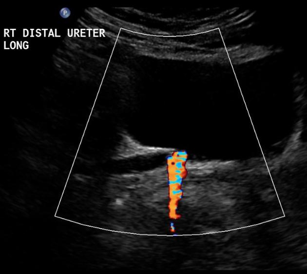
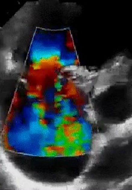
Color flow Doppler (ultrasound) The use of color flow Doppler (CFD) or color Doppler imaging (CDI) (or simply color Doppler) sonography allows the visualization of flow direction and velocity within a user defined area. A region of interest is defined by the sonographer, and the Doppler shifts of returning ultrasound waves within are color-coded based on average velocity and direction.
Physics
In a manner similar to pulsed wave Doppler (PWD), color flow Doppler utilizes intermittent sampling of ultrasound waves thereby avoiding the range ambiguity characteristic of continuous wave Doppler (CWD). Pulsed wave Doppler, however, is limited to the interrogation of flow velocity and direction along a single line at a certain depth (defined by the sample volume or gate); Color flow Doppler simultaneously interrogates multiple sample volumes (with each pixel representing a sample volume) along an array of scan lines 1. Information regarding the flow velocity and direction is arbitrarily color-coded and rendered onto a grey-scale (or M-mode) image. Flow that travels away from the transducer (negative Doppler shift) is depicted in blue, and flow that is traveling toward the transducer (positive Doppler shift) is depicted in red, with lighter shades of each color denoting higher velocities. A third color, usually green or yellow, is often used to denote areas of high flow turbulence. These colors are user-definable and may be reversed, however this is generally inadvisable as it may confuse later readers of the images. When the Nyquist limit is reached, however, aliasing results in apparent flow direction occurring in the opposite direction to actual flow, a limitation shared with pulsed wave Doppler. The velocity at which flow aliases is further impacted by the transducer used and the depth at which one is insonating flow.
Clinical use
Color flow Doppler is used frequently in sonography to semiquantitative overall blood flow to a region of interest. Depiction of the general velocity and direction of blood flow within the heart and blood vessels is of primary importance in echocardiography and =vascular-ultrasound&lang=us]vascular ultrasound respectively. It also allows the generation of unique phenomena such as the fluid color sign or the twinkling artifact and allows the targeting of spectral Doppler for a quantitative assessment of blood flow 2.
Techniques
There are several general parameters that need to be taken into account to obtain an optimum color flow Doppler scan: types of transducers, location and number of focal zones, depth of field, 2D gain setting, scan orientation, and image zoom, Specific color flow Doppler settings such as color and spectral parameters should also be optimized.5 A high 2D gain setting would suppress the color flow data while a low gain setting will highlight the color Doppler information.5 Increasing the depth of field would slow the frame rate while reducing the depth of field would increase the frame rate.5 Using multiple focal zones would reduce the frame rate, thus single focal zone is preferred for the color Doppler study to improve the frame rate.5 Setting a very high color velocity scale would show an apparent absence of flow while setting a very low color velocity scale would show aliasing of the flow signals.5
History and etymology
Named after Austrian physicist, Christian Andreas Doppler (1803-1853) 4.
عدد المساهمات : 2464نقاط : 4252السٌّمعَة : 9الجنس : علم بلدك : تاريخ الميلاد : 03/04/1950تاريخ التسجيل : 30/07/2012العمر : 74 الموقع : السودان - سنارالعمل/الترفيه : طبيب عمومى وموجات صوتيةالساعة الان : دعائي :
موضوع: Abbreviations السبت مايو 04, 2024 8:09 am
Abbreviations CW: Continuous wave/ PRF: Pulse repetition frequency/ PW Pulsed wave / SNR Signal-to-noise ratio/ Tx, Rx Transmit, receive/ One of the main advantages of ultrasound imaging compared to other imaging modalities (e.g., MRI and CT) is the ability to measure blood velocities in real time and at a high temporal resolution. This has made ultrasound imaging the main working tool for cardiovascular diagnosis, where conditions such as vessel stenosis, heart valve leakages, and left ventricle (LV) filling patterns can be quantitatively evaluated at bedside. Three modalities exist in current clinical systems, continuous-wave Doppler (CW Doppler), pulsed-wave Doppler (PW Doppler), and color Doppler imaging (CDI). The first two are also termed spectral Doppler (includes both CWD & PWD) as the complete blood velocity spectrum in a specific region is shown over time in a sonogram display. The latter (CDI) shows a 2D or 3D parametric image of the mean blood velocity and direction, which provides better information of the spatial blood velocity distribution and vessel geometry. The techniques are based on the principle of estimating the frequency shift (CW Doppler) or phase shift (PW Doppler and CDI) due to the axial movement of blood scatterers. Currently, all modalities are used in a clinical evaluation : typically CDI is used initially to get a qualitative overview of the blood velocity distribution and to detect regions of abnormal flow patterns.
CW or PW Doppler is further used for an in-depth evaluation and quantification of blood velocities at the specific sites of interest. The first clinical CW and PW Doppler systems appeared in the late 1960s and 1970s (Baker, 1970, Baker et al., 1964, Barber et al., 1974, Peronneau and Leger, 1969, Wells, 1969), while the clinical foundation rationalizing its use as a noninvasive diagnostic tool was established by the late 1970s and early 1980s (Brubakk et al., 1977, Fitzgerald and Drumm, 1977, Hatle et al., 1978, Holen et al., 1976, Johnson et al., 1973, McCallum et al., 1978). Commercial CDI systems were introduced in the mid-1980s (Eyer et al., 1981, Kasai et al., 1985) and were a major breakthrough in many clinical applications such as cardiology and fetal medicine. Since then, a continuous development has extended the functionality of ultrasound Doppler imaging, and the techniques are today used in a wide range of clinical applications. In the following chapters, a technical overview of current Doppler modalities is provided. Alternative approaches to blood velocity currently used mostly in research are further introduced, and future directions are given describing the latest advances in the research community. Similar approaches as those described in this chapter are used also to quantify tissue velocities, the so-called tissue Doppler imaging (TDI). This is in particular used in cardiac imaging to evaluate the contractile properties of the myocardium. This topic is only briefly covered in this chapter, and the reader is referred to other excellent textbooks such as Marwick et al. (2007) and Sutherland et al. (2006) for more information. Other textbooks describing ultrasound Doppler imaging may with benefit be used to achieve further understanding of specific clinical use or technical parts of ultrasound Doppler imaging, for instance, Evans and McDicken (2000), Jensen (1996), Angelsen and Torp (2000), Cobbold (2007), and Szabo (2004).
د.كمال سيد Admin
عدد المساهمات : 2464نقاط : 4252السٌّمعَة : 9الجنس : علم بلدك : تاريخ الميلاد : 03/04/1950تاريخ التسجيل : 30/07/2012العمر : 74 الموقع : السودان - سنارالعمل/الترفيه : طبيب عمومى وموجات صوتيةالساعة الان : دعائي :
موضوع: 32wk Doppler US السبت مايو 04, 2024 7:43 pm
Fetal doppler ultrasound is a common imaging technique in obstetrics to examine the fetal heart and blood vessels, especially important at 32 weeks. Not only is it safe and highly accurate, 32 weeks pregnant doppler ultrasound can also detect abnormalities that are often invisible with ultrasound. Compared with normal ultrasound, fetal doppler ultrasound has many outstanding advantages such as: The procedure is not complicated. Appropriate cost. Safe, non-invasive, no harm to mother and fetus. High accuracy, the ability to detect problems earlier and more accurately.
Indication for doppler ultrasound 32 weeks pregnant
The obstetrician will appoint 32 weeks pregnant Doppler ultrasound to: Assess 1/ the blood flow in parts of the fetus such as the umbilical cord, brain, heart to determine the ability of the fetus to absorb oxygen and nutrients . Investigation of 2/ blood flow through arterioles and veins. Thereby estimating fetal development. 3/Screening for diseases and birth defects of the fetus, giving remedial measures and dealing with them as soon as possible. 4/ Assess the appropriate time of delivery. Subjects assigned to 32 weeks pregnant doppler ultrasound include: Pregnant women with1/ multiple pregnancies. The fetus is affected by 2/ Rh antibodies due to the mother's Rh incompatibility. The fetus is affected by 3/ Parvovirus disease - an infectious disease caused by the Canine Parvovirus virus. Pregnant women with 4/ a history of (LBW) low birth weight babies. 5/ The fetus is growing slowly compared to the standard growth chart. Pregnant women with a 6/ history of late miscarriage or fetal death shortly after birth. Pregnant women with 7/ diseases during pregnancy such as hypertension, diabetes,... Pregnant women with 8/ low body mass index (BMI) or higher than standard. Pregnant women 9/ smoke during pregnancy. Thirty Two weeks pregnant doppler ultrasound is an important time that women should not ignore because the last 3 months of pregnancy is when there is a risk of morphological abnormalities in blood vessels, heart, brain. This technique helps to assess the health status and development of the fetus comprehensively and detect birth defects and abnormalities, if any, early. Thereby, your doctor will develop a plan to ensure the pregnancy has the best outcome for both you and your baby. https://www.vinmec.com/en/news/health-news/obstetrics-gynecology-and-assisted-reproductive-technologies-art/indications-for-doppler-ultrasound-at-32-weeks-gestation/
د.كمال سيد Admin
عدد المساهمات : 2464نقاط : 4252السٌّمعَة : 9الجنس : علم بلدك : تاريخ الميلاد : 03/04/1950تاريخ التسجيل : 30/07/2012العمر : 74 الموقع : السودان - سنارالعمل/الترفيه : طبيب عمومى وموجات صوتيةالساعة الان : دعائي :
موضوع: Doppler ultrasound pregnancy السبت مايو 04, 2024 7:53 pm
Doppler ultrasound uses sound waves to detect the movement of blood in vessels. It is used in pregnancy to study blood circulation in the baby, uterus and placenta. Using it in high‐risk pregnancies, where there is concern about the baby's condition, shows benefits. One of the main aims of routine antenatal care is to identify the 'at risk' fetus in order to apply clinical interventions which could result in reduced perinatal morbidity and mortality. Doppler ultrasound study of umbilical artery waveforms helps to identify the compromised fetus in 'high‐risk' pregnancies and, therefore, deserves assessment as a screening test in 'low‐risk' pregnancies. Growth‐restricted fetuses are at increased risk of mortality and morbidity (Bernstein 2000; Fisk 2001). The serious morbidity includes intraventricular haemorrhage, bronchopulmonary dysplasia, necrotising enterocolitis, infection, pulmonary haemorrhage, hypothermia and hypoglycaemia (Fisk 2001). Early antenatal detection, treatment where appropriate, and timely delivery could minimise the risks significantly. Different Doppler methods are used in obstetrics: continuous‐wave, (CWD) pulsed‐wave (PWD), colour (CD) and power Doppler flow (PD) (Eik‐Nes 1980; Mires 2000). Doppler ultrasound examination can be performed as a part of a more detailed ultrasound assessment that includes fetal biometry and anatomical survey, or as a separate ultrasound examination. Flow of the umbilical and fetal arteries is most often quantified either by pulsatility index (PI) or (RI) resistant index (Burns 1993; Nelson 1988). These indices reflect the down stream vascular resistance by quantifying the differences between the peak systolic (PSV) and the end‐diastolic velocity (EDV) within blood vessels of interest in each cardiac cycle. A high ratio in umbilical artery indicates a high vascular impedance and possible feto‐placental compromise. In extreme circumstance the blood flow at the end of diastole may be absent or even reversed. Initial Doppler studies have been restricted to the umbilical artery, but other fetal vessels have recently become a focus of interest including middle cerebral artery and ductus venosus. Although stillbirths and fetal complications related to placental problems are rare in uncomplicated pregnancy, the impact is devastating. Current methods for the assessment of fetal well‐being and detection of compromised fetus in the routine antenatal care include:1/ symphysis fundal height measurement from the 24th week (Neilson 1998a; NICE 2008), 2/ fetal movements charts (Mangesi 2007) and 3/ antenatal cardiotocography (Pattison 1999). None of them, however, have proven ability to make an impact on perinatal mortality and morbidity. AN: antenatal BP: blood pressure CI: confidence interval CTG: cardiotocography CS: caesarean section FD: fetal distress ITT: intention‐to‐treat IUGR: intrauterine growth restriction NICU: neonatal intensive care unit PIH: pregnancy‐induced hypertension PNM: perinatal mortality RR: risk ratio SCBU: special care baby unit SGA: small‐for‐gestational age US: ultrasound VS: versus https://www.ncbi.nlm.nih.gov/pmc/articles/PMC6464774/ Doppler ultrasound is a specialized test that uses sound waves to measure the blood flow through blood vessels. During pregnancy, Doppler Ultrasound is used to check 1/ the blood flow to and from the placenta, which is the organ that delivers oxygen and nutrients to your growing baby. Doppler ultrasound can be used to 2/ detect any problems that may be affecting your baby's growth and development. For example, if the blood flowto the placenta is reduced, it could lead to a condition called placental insufficiency or fetal growth restriction, which can lead to slow growth of the baby. Doppler ultrasound is 3/ part of every pregnancy scan. It also allows for 4/ early detection of problems, allowing for timely intervention and care. For example, during the first trimester, if the Uterine artery Doppler is abnormal, doctors will advise blood thinners to improve the blood flow across the placenta. In your third trimester, a Doppler ultrasound study will help the doctor to 5/ plan the delivery. So make sure the Doppler study is included in your routine Pregnancy scans.
د.كمال سيد Admin
عدد المساهمات : 2464نقاط : 4252السٌّمعَة : 9الجنس : علم بلدك : تاريخ الميلاد : 03/04/1950تاريخ التسجيل : 30/07/2012العمر : 74 الموقع : السودان - سنارالعمل/الترفيه : طبيب عمومى وموجات صوتيةالساعة الان : دعائي :
موضوع: رد: Doppler US الأحد مايو 05, 2024 11:56 am
د.كمال سيد Admin
عدد المساهمات : 2464نقاط : 4252السٌّمعَة : 9الجنس : علم بلدك : تاريخ الميلاد : 03/04/1950تاريخ التسجيل : 30/07/2012العمر : 74 الموقع : السودان - سنارالعمل/الترفيه : طبيب عمومى وموجات صوتيةالساعة الان : دعائي :
موضوع: Doppler ultrasound in pregnancy الأحد مايو 05, 2024 12:02 pm
Doppler scan 1/ measures blood flow using sound waves, aiding in 2/ detecting vascular issues or monitoring fetal 3/ well being during pregnancy. It is safe when done by trained professionals, & lasts only a few minutes. why indicated ? If a doctor finds any anomaly during normal US scans, a Doppler scan is done for further investigation. Doppler scan is done to check : 1/ the placental blood flow 2/ the fetal umbilical blood flow 3/ the blood flow in the heart & brain 4/ If the mother is carrying twins or more 5/ If the mother has high or low BMI 6/ If there are medical conditions like DM or high blood presure 7/ If the baby is affected by Rhesus antibodies 8/ the baby's growth rate Dr will ask for a Doppler test in the following cases : 1/ multiple pregnancies 2/ placenta problems 3/ to determine the health condition of the mother 4/ to determine the health condition of the fetus how Doppler test in pregnancy done ? 1/ A gel is applied on the belly for a better contact, ensuring clear sound wave transmission 2/ the scan is painless & quick & provides real time images for analysis
CardioTocoGraphy (CTG)1/ measures baby's heart rate. At the same time it also 2/ monitors uterinecontractions. CTG is used both before birth (antenatally) and during labour, to 3/ monitor the baby for any signs of distress.
د.كمال سيد Admin
عدد المساهمات : 2464نقاط : 4252السٌّمعَة : 9الجنس : علم بلدك : تاريخ الميلاد : 03/04/1950تاريخ التسجيل : 30/07/2012العمر : 74 الموقع : السودان - سنارالعمل/الترفيه : طبيب عمومى وموجات صوتيةالساعة الان : دعائي :
موضوع: doppler physics الأحد مايو 05, 2024 1:07 pm
What is the principle of ultrasound? An electric current passes through a cable to the transducer and is applied to the crystals, causing them to deform and vibrate. This vibration produces the ultrasound beam. The frequency of the ultrasound waves produced is predeterminedby the crystals in the transducer. [rtl]•Correct interpretation of flow images requires knowledge of physical and technical factors that influence the Doppler signal. [/rtl] [rtl]•Artefacts caused by 1/physical limitations of the modality or 2/ inappropriate equipment settings may result in displayed flow conditions that may differ considerably from the actual physiological situation. [/rtl] [rtl]•As a consequence, artefacts in Doppler imaging may be confusing and lead to misinterpretation of flow information.[/rtl] [rtl]•DOPPLER SIGNAL •The Doppler effect is a change in wavelength (frequency) of sound resulting from motion of a source, receiver or reflector. As the US TXR is a stationary source and receiver, the Doppler effect arises from reflectors in motion—for all practical purposes these are the erythrocytes. • When a pulse is reflected from erythrocytes, the frequency of the wave received differs from that, which is transmitted. •This difference is known as the Doppler shift, named after the Austrian physicist and mathematician Chr. Andreas Doppler, who first described the phenomenon for light in 1843.5 there are two succsessive 2 doppler shifts involved: •1/ First, the sound from the stationary transmitting TXR is received by the moving erythrocytes. •2/Second, the erythrocytes act as moving sources as they •re-eradiate the US back toward the transducer, which is now the stationary receiver. •These two Doppler shifts account for factor 2 in the Doppler equation : •fd = ft – fr = 2 ft v cos^(ceta) / C DOPPLER EQUATION : •fd = ft – fr = 2 ft v cos^(ceta) / C •where: fD is the Doppler shift, ft is the transmitted frequency, fr is the received frequency, v is the blood velocity, θ (ceta) is the insonation angle (the angle between the US beam and the blood flow), and c is the speed of sound. •The Doppler shift is thus directly proportional to the velocity of the flow, (v),to cosine of the insonation angle, θ, and to the transmitted frequency of the US, ( ft).6 (vct) •Angle of insonation is defined as the angle of the ultrasound beam relative to the tissue or organ of interest. The strongest echoes are produced when the angles of incidenceapproach the angle of reflection. • The Greek letter θ (theta) is used in math as a variable to represent a measured angle. For example, the symbol theta appears in the three main trigonometric functions: sine, cosine, and tangent as the input variable. •PULSED DOPPLER •The Doppler circuitry determines the change (shift) in frequency indirectly. With a series of pulses, the phase of the returning signals are compared with the phase of the emitted signal. •A change in phase translates to a change in frequency; eg, when the returning signal is compared with the emitted one, wave tops will not meet wave tops because the distance between wave tops has changed. The number of these pulses per second is called the pulse repetition frequency (PRF) •Insonation angle, (Doppler angle) •This is the angle between the path of the Doppler pulses (TXR) and the direction of flowin the vessel. •When this angle is 90°, there will be no frequency shift as can be seen from the equation above: cos(90°) = 0. •The maximum frequency shift of a given vessel is obtained when the direction of flow matches the direction of the Doppler pulses (Doppler angle = 0, flow directly towards or away from the transducer). •Blood velocity versus Doppler shift •The Doppler circuitry determines the change in frequency and, this may only be translated into a blood velocity if the insonation angle is recorded and is included in the calculation. Nevertheless, all newer equipment report blood velocities (both in spectral Doppler and on the colour bar) assuming that the Doppler angle is zero. •This is, however, more often wrong than right and we are in fact dealing with frequency information unless angle correction has been performed (the process of informing the machine of the insonation angle).•Angle correction is only possible in spectral Doppler and is not an issue in a rheumatological setting. •COLOUR DOPPLER, VELOCITY DOPPLER AND COLOUR FLOW MAPPING •In color flow Doppler (CFD), real-time presentation of flow information in colour is superimposed on the grey-scale morphological image. •In colour Doppler, analysis is performed in the colour box, which is divided into cellsy. • Each cell behaves like an independent Doppler gate with its own Doppler analysis. •The mean frequency shift for each cell is computed and displayed as a colour. •The colours that arise from the detected Doppler shifts primarily indicate qualitative direction of flow. •Generally, red is used to indicate a flow towards the transducer and blue away from the transducer. •Different hues of red (or blue) indicate different velocities (in reality different frequency shifts). •Lighter hues are used to indicate higher frequency shifts. •Interpreting the colours •The Doppler circuitry merely detects movements up and down in the image plane. •A dark red spot may therefore be blood moving slowly, directly towards the transducer or blood moving quickly at an angle close to 90°. •As we generally do not know the insonation angle to the vessels we cannot compare velocities between vessels. •POWER DOPPLER (ENERGY DOPPLER) •PDdisplays power of the Doppler shift in each cell instead of the mean frequency shift. This gives PD a theoretical advantage over colour Doppler with regard to sensitivity. Disregarding direction of flow (negative or positive frequency shift) and disregarding velocity (high orlow frequency shift) the power (energy) of the many different frequency shifts inside a cell are added to form the power signal. •The power mode does not measure velocity or directionand is very sensitive to flow. Therefore, it is almost angle independent and without aliasing.7 ••The wall filter in ultrasound is a way of filtering out low or high frequency Doppler signals. ... •In clinical ultrasound, it is usually used to filter out very low frequencies that may add noise to a spectral Doppler waveform •A typical use is removing the low frequency reverberation of an arterial wall. • DOPPLER PARAMETERS •The most important adjustable parameters are : 1/ Doppler frequency, Doppler 2/ gain, 3/ PRF, 4/ colour priority, 5/ filter, 6/ focus, p7/ Persistence, 8/ colour box position and size. •Patient positioning and scanning technique further influence the quality of the Doppler examination. •Doppler frequency •A lower Doppler frequency will allow more penetration but also a more grainy Doppler image (larger colour pixels). Thus, higher Doppler frequency gives a more detailed image of the vessels but at the expense of penetration. •The trade-off between penetration and sensitivity is somewhat unpredictable and resolution is in this context really not an issue. •The ability to depict slow flow in a small vessel (with a weak Doppler reflection) is enhanced by a lower frequency (because the weak reflection has more penetration) but is also enhanced by a higher frequency because the Doppler shift is higher (if the reflection is powerful enough to penetrate). •The optimum frequency must be found in practice and not in theory. •Colour box •The numerous Doppler analyses inside the colour box are quite demanding on computing power of the US unit. •The frame rate goes down when colour is added, and in order to obtain as live an image as possible (high frame rate) it is therefore generally recommended to make the box as small as possible. •We do, however, recommend always letting the box go to the top of the image in order to be aware of reverberation artefacts(see reverberations).
د.كمال سيد Admin
عدد المساهمات : 2464نقاط : 4252السٌّمعَة : 9الجنس : علم بلدك : تاريخ الميلاد : 03/04/1950تاريخ التسجيل : 30/07/2012العمر : 74 الموقع : السودان - سنارالعمل/الترفيه : طبيب عمومى وموجات صوتيةالساعة الان : دعائي :
موضوع: رد: Doppler US الأحد مايو 05, 2024 2:10 pm
[rtl]•COLOUR DOPPLER, VELOCITY DOPPLER AND COLOUR FLOW MAPPING[/rtl] [rtl]Color Doppler (CD) test uses a computer to change sound wave measurements into different colors. The colors show the speed and direction of blood flow.[/rtl] [rtl]The speed of the blood flow is shown with a color scale. Usually, blood flow away from the probe is shown in blue, while blood flow toward the probe is red.[/rtl] [rtl]Doppler ultrasound is a noninvasive test that can be used to measure the blood flow through your blood vessels. It works by bouncing high-frequency sound wavesoff red blood cells that are circulating in the bloodstream. A regular ultrasound uses sound waves to produce images, but can't show blood flow.[/rtl] [rtl] Normal doppler US test resuls mean that 1/ the blood vessels show NO signs of narrowing, clots, or closure, 2/ and the arteries have normal blood flow.[/rtl] [rtl]Color coded Doppler flow mapping (color flow mapping or color Doppler) is a newly developed technology which displays blood flow as color-coded information on the two-dimensional image. This method is an application of the pulsed Doppler method.[/rtl] [rtl]Colour flow mapping (CFM) produces a two-dimensional representation of blood flow within the heart and great vessels by analysing data acquired from multiple pulsed Doppler sample volumes, and displaying mean velocity and turbulence of flow at each site.[/rtl] [rtl]How to read a color Doppler report ? Velocities recorded in a sample volume of the pulsed wave Doppler can be presented with a color. A color scale from blue to red is conventionally used. Blue color implies velocities (movement) away from the transducer and red color implies velocities (movement) towards the transducer.[/rtl] [rtl]What is color scale in Doppler ?[/rtl] [rtl]In color Doppler mode, color assignments (job, function) are set and displayed as a 'map' on the screen. Traditionally, red is the color assigned to flow towards the transducer, and blue is assigned to flow away from the transducer.[/rtl] [rtl]Color Doppler converts the blood flow measurements into an array of colors to help show the speed and direction of blood flow through the vessel. Power Doppler is even more sensitive than color Doppler in detecting very slow blood flow, although it does not provide information about the direction of blood flow.[/rtl] [rtl]Color maps are used to display a single-band raster consistently with the same colors. Each pixel value is associated with a color, defined as a set of red, green, and blue (RGB)[/rtl] [rtl]The Colormap function is a raster data renderer. It transforms the pixel values to display the raster data as either a grayscale or an RGB color image based on a color scheme or specific colors in a color map file.[/rtl] [rtl]Colour flow image obtained by superimposing Doppler information on the pulse-echo image. The subject's head is to the left of the scan, and so the arterial flow is from left to right and the venous flow from right to left.[/rtl] [rtl]Color Doppler imaging parameters, including packet size, color gate, color gain, color reject, capture function, velocity map, and velocity tag, are described. Color Doppler image quality is characterized by motion discrimination, temporal resolution, spatial resolution, and uniformity.[/rtl] [rtl]what is the principle of Doppler ?[/rtl] [rtl]Doppler effect in physics is defined as the increase (or decrease) in the frequency of sound, light, or other waves as the source and observer move towards (or away from) each other. Waves emitted by a source travelling towards an observer get compressed.[/rtl] [rtl]Doppler waveforms refer to the morphology of pulsatile blood flow velocity tracings on spectral Doppler ultrasound. Waveformsdiffer by the vascular bed (peripheral, cerebrovascular, and visceral circulations) and the presence of disease.[/rtl] [rtl]Diagnostic Performance of Color Doppler Sonography According to Population Sex. Color Doppler sonography showed a sensitivity of 81.8% for men and 61.8% for the whole female population including pregnant women (p < 0.001) and a specificity of 97.7% for men and 96.9% for women.[/rtl] [rtl]what is Doppler score ?[/rtl] [rtl]The blood flow was assessed by a color Doppler score based on the intensity of the color signal with the following value ranges: (1) no flow, (2) minimal flow, (3) moderate flow, and (4) high vascular flow.[/rtl] [rtl]What is color Doppler scale?[/rtl] [rtl]Color Doppler US analyzes phase, frequency, and amplitude information encoded in the returning echoes to overlay a color image displaying blood flow onto a real-time gray-scale image. Color is assigned to objects that are in motion (e.g., red blood cells in flowing blood) based on the direction of the phase shift.[/rtl] [rtl]The Importance of Colormaps. Colormaps control the way data are understood when plotted because they map data values to color. When the colors used do not match the way humans perceive color, there can be a mismatch between interpretation and the data.[/rtl] [rtl]•In CFD, real-time presentation of flow information in colour is superimposed on the grey-scale morphological image.[/rtl] [rtl]•In colour Doppler, analysis is performed in the colour box, which is divided into cells.[/rtl] [rtl]• Each cell behaves like an independent Doppler gate with its own Doppler analysis. [/rtl]
د.كمال سيد Admin
عدد المساهمات : 2464نقاط : 4252السٌّمعَة : 9الجنس : علم بلدك : تاريخ الميلاد : 03/04/1950تاريخ التسجيل : 30/07/2012العمر : 74 الموقع : السودان - سنارالعمل/الترفيه : طبيب عمومى وموجات صوتيةالساعة الان : دعائي :
موضوع: رد: Doppler US الأحد مايو 05, 2024 5:51 pm
Principles of Color Doppler COLOR BOX The colors represent the speed and direction of blood flow within a certain area of the image (color box). The color box is divided into small sample regions (one color pixel). Each of these represent the mean velocity within the region as measured by multiple PW sample volumes. The mean velocity is then converted into a specific color. By definition, flow towards the transducer is depicted in red while flow away from the transducer is shown in blue. Different shades of red and blue are used to display velocity. Lighter shades of color are assigned to higher velocities. The exact manner in which velocities are displayed is defined by the so-called color map. Ultrasound scanners are usually provided with several maps from which the investigator may choose those he/she prefers. Some maps encode turbulent flow in yellow or green. Turbulent flow is present when there are large variations in flow velocity within the sample region. The information is consistently updated from frame to frame, thus providing a clear view of how blood "flows". The main advantage of color Doppler (CD) is the fact that one can view blood flow simultaneously in many regions of the heart. However, in contrast to PW and CW Doppler, color Doppler only permits semiquantitative assessment of blood flow velocities. Since color Doppler (CD) is based on the same principles as PW Doppler it is also subject toaliasing. When the velocity of blood flow exceeds the Nyquist limit (0.4-0.7 m/s) it becomes impossible to measure the direction of blood flow. This condition results in an abrupt shift of color from red to blue or vice versa. Note that the brightest shades of red and blue lie adjacent to each other at the aliasing point. This accentuates regions of aliased flow for the eye and helps the investigator to detect high-velocity jets. When the Nyquist limit is reached the color changes abruptly (red to blue, or blue to red). As variations in velocity are quite marked in regions of turbulent flow, you will find a "mosaic" pattern of colors. The phenomenon of aliasing and "mosaic colors" provides a good delineation of jets (and measurement of PISA) The Proximal Isovelocity Surface Area (PISA) measurement, also known as the "flow convergence" method, can be used in echocardiography to estimate the area of an orifice through which blood flows. As with PW Doppler, the Nyquist limit (the velocity at which aliasing occurs) also depends on the imaging depth and the ultrasound frequency one is using. The Nyquist limit can be adjusted (within a certain range) by changing the pulse repetition frequency (PRF). https://123sonography.com/ebook/principles-color-doppler
عدل سابقا من قبل د.كمال سيد في الإثنين مايو 13, 2024 8:44 am عدل 1 مرات
د.كمال سيد Admin
عدد المساهمات : 2464نقاط : 4252السٌّمعَة : 9الجنس : علم بلدك : تاريخ الميلاد : 03/04/1950تاريخ التسجيل : 30/07/2012العمر : 74 الموقع : السودان - سنارالعمل/الترفيه : طبيب عمومى وموجات صوتيةالساعة الان : دعائي :
موضوع: رد: Doppler US الأحد مايو 05, 2024 7:26 pm
M-Mode ultrasound imaging The M-mode was the preferred imaging modality in the early days of ultrasound. M-mode is defined as time motion display of the ultrasound wavealong a chosen ultrasound line. It provides a monodimensional view of the heart. All of the reflectors along this line are displayed along the time axis. The advantage of the M-mode is its very high sampling rate, which results in a high time resolution so that even very rapid motions can be recorded, displayed, and measured. The disadvantage is that the US line is fixed to the tip of the US sector. It may therefore be difficult to align the M-mode perpendicular to the structures which are displayed (i.e. the septum), thus leading to false measurements. Anatomical M-mode circumvents this limitation by reconstructing the M-mode from the 2D image (post-processing). The anatomical M-mode permits free positioning of the cursor line. However, the time resolution is significantly less than that of the conventional M-mode. Mode tracing across the aortic sinus and the left atrium/M-mode tracing across the left ventricle The M-mode can be combined with other imaging modalities such as color or tissue Doppler. Curved M-mode is a color display format in which functional information (such as velocities, strain, strain rate) concerning different segments of the heart (such as the 4-chamber view) are displayed along an M-mode line which follows the myocardial walls. The M-mode line "curves" around the myocardium. Starting at the basal inferior segment it moves to the apex and back to the basal lateral wall. The functional information is color encoded. Tissue Doppler Mode/Color Doppler M-mode The M-mode has lost much of its importance but is still valuable in certain situations. Advantages:
High time resolution
Allows timing of intervals
M-mode — When is it used?
Aorta/ left atrium (measurements, opening of aortic valve)
Mitral/Prosthetic valve (type of valve)
Tricuspid annular plane systolic excursion (TAPSE) for RV function
Left/right ventricle (measurements, LV function)
Endocarditis (motion of suspected vegetation)
Mitral valve (Mitral stenosis)
https://123sonography.com/content/17-m-mode Continuous-wave Doppler ultrasound Continuous-wave Doppler (CWD) utilizes continuous transmission and reception of ultrasound waves. This is accomplished by two dedicated transducer elements: one that solely sends a signal and another that only receives. As no pulses are emitted, CW Doppler does not permit us to determine where the wave is reflected. It is not site specific. We only know that the depicted velocity curve stems from a reflector somewhere along the path of the ultrasound beam, which is known as the CW Doppler line. The advantage of CW Doppler is its ability to measure high velocities, which are frequently observed in pathologies of the heart (e.g. aortic stenosis) CW Doppler spectrum of tricuspid regurgitation What information one can obtained with spectral Doppler Spectral Doppler provides information about the velocity of blood flow over time. Therefore, it permits measurement of maximal and mean velocity. Maximal velocity is equivalent to the highest point of the curve (below or above the zero line). Mean velocity, as the term denotes, is calculated as the mean of all velocities. This is done by simply tracing the curve and integrating the velocities below the curve (don't worry - the machine does it for you). Doppler curves also provide valuable information about the timing of events, especially when relating the Doppler curves to the ECG tracing. For instance, we are able to measure time intervals such as the ejection period, isovolumetric relaxation time, or diastolic filling time. By viewing the slope of the curves (i.e. deceleration and acceleration times), we can determine how quickly the velocity changes. Velocities are also related to "pressures". To understand this we will have to first discuss a physical principle that was first described by the Swiincreased before the point of obstructionss physicist Bernoulli in the 18th century : When a fluid passes through a narrow space, its flow velocity increases. This is similar to a river with rapids in those regions in which the river bed is narrow. Pressure is increasedbefore the point of obstruction and is low behind the obstruction. The difference between these pressures is known as the pressure gradient. The pressure gradient is actually the driving force behind the increase in velocity. Bernoulli found that velocity increases exponentially (tremrdousely) with the pressure gradient. With the aid of Bernoulli's equation one can calculate the pressure gradient from the velocity. As pressure gradients are also related to the degree of obstruction, we can apply this principle to quantify lesions such as aortic stenosis or shunts. Bernoulli's equation is actually quite complex. It considers factors such as viscosity and flow coefficients. However, the simplified version of Bernoulli's equation is adequate for the purpose of medical ultrasound. simplified Bernoulli's equation The simplified version of Bernoulli's equation permits easy estimation of pressure gradients from velocities. Bernoulli's equation is the most important formula in echocardiography. As you will see later, it will be used not only to derive maximum and mean pressure gradients but also for many other calculations. https://123sonography.com/book/395
عدل سابقا من قبل د.كمال سيد في الإثنين مايو 13, 2024 8:50 am عدل 1 مرات
د.كمال سيد Admin
عدد المساهمات : 2464نقاط : 4252السٌّمعَة : 9الجنس : علم بلدك : تاريخ الميلاد : 03/04/1950تاريخ التسجيل : 30/07/2012العمر : 74 الموقع : السودان - سنارالعمل/الترفيه : طبيب عمومى وموجات صوتيةالساعة الان : دعائي :
موضوع: رد: Doppler US الإثنين مايو 06, 2024 9:51 am
[rtl]•COLOUR DOPPLER, VELOCITY DOPPLER AND COLOUR FLOW MAPPING[/rtl] [rtl]•In CFD, real-time presentation of flow information in colour is superimposed on the grey-scale morphological image.[/rtl] [rtl]•In colour Doppler, analysis is performed in the colour box, which is divided into cells.[/rtl] [rtl]• Each cell behaves like an independent Doppler gate with its own Doppler analysis. [/rtl] [rtl]•The mean frequency shift for each cell is computed and displayed as a colour. •The colours that arise from the detected Doppler shifts primarily indicate qualitative direction of flow. •Generally, red is used to indicate a flow towards the transducer and blue away from the transducer. •Different hues of red (or blue) indicate different velocities (in reality different frequency shifts). •Lighter hues are used to indicate higher frequency shifts. •Interpreting the colours •The Doppler circuitry merely detects movements up and down in the image plane. •A dark red spot may therefore be blood moving slowly, directly towards the transducer or blood moving quickly at an angle close to 90°. •As we generally do not know the insonation angle to the vessels we cannot compare velocities between vessels. •POWER DOPPLER (ENERGY DOPPLER) •PD displays power of the Doppler shift in each cell instead of the mean frequency shift. This gives PD a theoretical advantage over colour Doppler with regard to sensitivity. Disregarding direction of flow (negative or positive frequency shift) and disregarding velocity (high or low frequency shift) the power (energy) of the many different frequency shifts inside a cell are added to form the power signal. •The power mode does not measure velocity or direction and is very sensitive to flow. Therefore, it is almost angle independent and without aliasing.7 •The wall filter in ultrasound is a way of filtering out low or high frequency Doppler signals. ... •In clinical ultrasound, it is usually used to filter out very low frequencies that may add noise to a spectral Doppler waveform. •A typical use is removing the low frequency reverberation of an arterial wall. • DOPPLER PARAMETERS •The most important adjustable parameters are 1/ Doppler frequency, 2/ Doppler gain, 3/ PRF, 4/ colour priority, 5/ filter, 6/ focus, 7/ persistence, 8/ colour box position and size. •Patient positioning and scanning technique further influence the quality of the Doppler examination. •Doppler frequency •A lower Doppler frequency will allow more penetration but also a more grainy Doppler image (larger colour pixels). Thus, higher Doppler frequency gives a more detailed image of the vessels but at the expense of penetration (less penetration). •The trade-off between penetration and sensitivity is somewhat unpredictable and resolution is in this context really not an issue. •The ability to depict slow flow in a small vessel (with a weak Doppler reflection) is enhanced by a lower frequency (because the weak reflection has more penetration) but is also enhanced by a higher frequency because the Doppler shift is higher (if the reflection is powerful enough to penetrate). •The optimum frequency must be found in practice and not in theory. •Colour box •The numerous Doppler analyses inside the colour box are quite demanding on computing power of the US unit. •The frame rate goes down when colour is added and in order to obtain as live an image as possible (high frame rate) it is therefore generally recommended to make the box as small as possible. •We do, however, recommend always letting the box go to the top of the image in order to be aware of reverberation artefacts(see reverberations). •Scale and pulse repetition frequency •PRF is the Doppler sampling frequency of the transducer and is reported in Hz. •The maximum Doppler shift frequency that can be sampled without aliasing is PRF/2, which is called the Nyquist limit.8 •The Nyquist limit may be presented on-screen as a blood velocity (the maximum measurable velocity of blood moving directly towards or away from the transducer) or in Hz (maximum measurable Doppler shift). •If the blood velocity is above the Nyquist limit, the machine will misinterpret the velocity and aliasing will occur. This is not an issue with PD. •The sensitivity of both colour Doppler (CD) and (PD) is affected by PRF adjustments. When a high PRF is chosen it is assumed that the investigator is interested in high velocities, and therefore filters that remove low flow to remove noise are applied (so-called linked controls). •Selecting a high PRF therefore makes the system insensitive to lower velocities because of the linked controls. •Colour priority (threshold) •When colour information is obtained, grey-scale information will often also be present and the machine has to decide whether to show one or the other. •Colour priority is a function that makes this decision for the machine. This function allows valid grey-scale information to override false Doppler information, eg, it helps suppress motion artefacts in the relatively hyperechoic tissue surrounding a pulsating artery (above a certain grey level, grey overrides colour). •This function also allows supposedly valid Doppler information to override false grey-scale information, eg, inside vessels colour overrides the relatively weak grey-scale reverberation artefacts (below a certain grey level colour overrides grey). This function explains why some Doppler artefacts apparently prefer to appear in dark regions of the image. It also explains why grey-scale gain may influence the amount of colour in the image (increasing grey-scale gain may result in more grey information being above the threshold where colour is suppressed). • Filters •Every Doppler instrument has high-pass filters, which eliminate the lowest Doppler shifts from the display. •The Doppler shifts originate from motion of the vessel wall and solid tissue. •These unwanted shifts are referred to as clutter or motion artefacts. •The filters—also called wall filters—may, however, eliminate signals from low velocity flow as the filters separate by frequency alone.910 •The PRF and wall filter are linked controls—the lowest possible wall filter is lower for a low PRF than a high. •The filters should be kept at their lowest setting for use in rheumatology. •Gain •The Doppler gain is independent of grey-scale gain. •The gain setting determines the sensitivity of the system to flow. •By lowering the gain, noise and motion artefacts may be prevented but weak flow signals will go undetected. •11 A gain setting that is too high results in random noise.1 •It is appropriate to set the Doppler gain by turning it up until random noise is encountered and then lowering it until the noise disappears.11 •Persistence •Persistence is a function that averagescolour information over a number of frames. •Most brochure images are made with maximum persistence because this results in all colour information over time being displayed in one image. •All vessels are then filled with colour. The dynamic nature of flow is, however, lost. •With low or no persistence the high or low resistance nature of arterial flow may be seen as blinking colour foci or colour foci with pulsating intensity, respectively. •In a rheumatological setting there is no advantage with high persistence Patient positioning •The patient must be positioned comfortably, and the area under investigation must be completely relaxed; otherwise, tension in the muscles and tendons will produce slight tremor resulting in movement artefacts. When scanning the hand and elbow regions, the arm should not rest on the abdomen or thorax because of respiratory movement. Some patients should also keep quiet during the Doppler examination because the voice itself may produce movement artefacts and because some patients cannot talk without involuntary movement of the hands. •Scanning technique •Most important in Doppler examinations is that very little pressure should be applied by the transducer. •The pressure will affect the haemodynamics with resulting decreased flow. •The use of generous amounts of scanning gel with visible gel between the transducer and skin will ensure light pressure. •Focusing •The Doppler uses the same focus point as the grey-scale image. In the focal zone, the pulse is most narrow and the pulse therefore has higher spatial peak energy. •As a consequence, the echoes generated in the focal zone have higher amplitudes. •It is therefore not surprising that the Doppler is very dependent upon focus positioning .•Pressure False findings of absence of flow may occur if the examiner presses too hard on the tissue with the transducer, thereby blocking the flow. •When scanning a concave or convex surface it may be tempting to press the surface flat with the transducer. Instead, a generous amount of scanning gel should be used, which obviates the need for pressure to obtain good acoustic contact. •
[/rtl]
عدل سابقا من قبل د.كمال سيد في الإثنين مايو 13, 2024 8:55 am عدل 1 مرات


 الثلاثاء أبريل 30, 2024 4:33 pm