
موقع د. كمال سيد الدراوي
طبي_ اكاديمي _ ثقافي _ تعليمي _ _ استشارات طبية_فيديو طبي
|
| | | كل شئ عن الأشعه السينيه |  |
| | | كاتب الموضوع | رسالة |
|---|
د.كمال سيد
Admin


عدد المساهمات : 2471
نقاط : 4259
السٌّمعَة : 9
الجنس : 
علم بلدك : 
تاريخ الميلاد : 03/04/1950
تاريخ التسجيل : 30/07/2012
العمر : 74
الموقع : السودان - سنار
العمل/الترفيه : طبيب عمومى وموجات صوتية
الساعة الان :
دعائي : 
|  موضوع: كل شئ عن الأشعه السينيه موضوع: كل شئ عن الأشعه السينيه  الثلاثاء أبريل 30, 2019 6:14 pm الثلاثاء أبريل 30, 2019 6:14 pm | |
| مقدمة في الأشعه السينيه
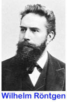
Wilhelm Conrad Röntgen was a German mechanical engineer and physicist, who, on 8 November 1895, produced and detected electromagnetic radiation in a wavelength range known as X-rays or Röntgen rays, an achievement that earned him the first Nobel Prize in Physics in 1901.
مع تقدم علوم طب الأسنان بدأت الأشعة تشغل مكانها المناسب إما بكونها اختصاصاً قائماً بحد ذاته، أو بكونها علماً متمماً لاختصاص آخر بحيث أن الأشعة تقدم وسيلة تشخيصية لا يمكن إهمالها.
منذ اكتشاف روتنجن للأشعة السينية عام 1895 ومحاولات الأطباء والأبحاث تتزايد يوماً بعد يوم للاستفادة القصوى من هذا الاكتشاف العظيم. ولكي يكون طبيب الأسنان قادر على فهم المظاهر الشعاعية الطبيعية والمرضية يجب أن يكون ملماً بشكل جيد بمعظم علوم طب الأسنان الأخرى وبالذات التشريح الوصفي والتشريح المرضي وبعض مبادئ علم الفيزياء.
تعريف الأشعة السينية وخواصها :
الأشعة السينية أو أشعة اكس (X-rays): هي جزء من الطيف الالكترومغناطيسي (electromagnetic spectrum) والتي تمتلك طول موجة تتراوح بين (0.01 – 10) نانو متر، فهي أقصر من طول موجة الطيف المرئي ولكنها في المقابل تمتلك توتر عالي (3×10¹⁶ – 3×10¹⁹ ) منه، وهذا ما يضعها في المنطقة بين الأشعة فوق البنفسجية (ultraviolet light) وأشعة غاما (gamma rays). ويمكن تمييزها عن أشعة غاما بطول موجتها أو تواترها بالإضافة إلى أنّ الأشعة السينية تصدر عن الالكترونات أما أشعة غاما فهي ناتجة عن النواة الذرية.
أول من درس هذا النوع من الأشعة هو العالم الألماني ويليام رونتغن (Wilhelm Röntgen) عام (1895) ولكنه ليس أول من اكتشفها فقد تم اصدار هذه الاشعة من أنبوب كروكس (Crookes tubes) الذي تم اختراعه سنة (1875). وقد سميت حينها بأشعة اكس ثم سميت بأشعة رونتغن وفي الوقت الحالي فيمكن استخدام إحدى التسميتين (وفي اللغة العربية سميت بالأشعة السينية كتعريب لأشعة اكس)
أنواع الأشعة السينية:
هنالك نوعين من الأشعة السينية (العالية والمنخفضة). وبشكل عام تمتلك الأشعة السينية طاقة تتراوح بين (100 eV) (الكترون فولط) و (100 keV) (كيلوالكترون فولط). فالأشعة التي تمتلك طاقة أعلى من (5-10 keV) تسمى بالأشعة السينية العالية والأشعة ذات الطاقة الأخفض سميت بالأشعة السينية المنخفضة. الأطوال الموجية للأشعة السينية العالية يمكن مقارنتها بالأبعاد الذرية ولذلك فهي تمتلك طاقة قادرة على اختراق المواد. أما الأشعة السينية المنخفضة يمكن أن تتلاشى وتمتص في الهواء أو يمكن أن تخترق الماء والأبعاد حتى 1 ميكرومتر.
مصادر الأشعة السينية:
تصدر الأشعة السينية عندما تضرب جسيمات تمتلك طاقة معينة سطح المادة. كما ويستخدم التسارع الالكتروني في إنتاج الأشعة السينية في أنبوب الأشعة السينية (وهو عبارة عن أنبوب مفرغ من الهواء يحتوي على كاتود (صفيحة أو سلك ذو شحنة موجبة) ومعدن يتلقى الشحنة الناتجة عن الكاتود). كما يمكن استخدام البروتونات أو أي شحنات موجبة. وهناك مصادر أخرى طبيعية للأشعة السينية منها غاز الرادون و النظائر المشعة أو الأشعة الآتية من الفضاء.
كيف تتأثر المادة بالأشعة السينية:
تمتلك الأشعة السينية طاقة معينة يمكن أن تؤثر بطاقة الروابط بين الذرات في الجزيئات وعلى هذا فإنّ التأثير يتعلق بقوة الارتباط بين الذرات وبطاقة الأشعة المؤثرة ولا علاقة للخواص الكيميائية للمادة على قوة تأثير أشعة اكس أو ضعفه.
متابعة الموضوع | |
| | | | د.كمال سيد
Admin
عدد المساهمات : 2471
نقاط : 4259
السٌّمعَة : 9
الجنس :
علم بلدك :
تاريخ الميلاد : 03/04/1950
تاريخ التسجيل : 30/07/2012
العمر : 74
الموقع : السودان - سنار
العمل/الترفيه : طبيب عمومى وموجات صوتية
الساعة الان :
دعائي :
| | موضوع: رد: كل شئ عن الأشعه السينيه الثلاثاء أبريل 30, 2019 6:14 pm | |
| استخدامات الأشعة السينية:
أغلب الناس تعرف الأشعة السينية بناء على معرفتهم باستخدامها في التصوير الطبي (تصوير الكسور وغيرها) ولكن هنالك العديد من الاستخدامات الأخرى:
في التشخيص الطبي، تستخدم في رؤية بنية العظام وشكلها ويستخدم فيها الأشعة السينية العالية. فالكتلة الذرية العالية للكالسيوم في الأسنان والعظام تمتص أشعة اكس بينما تخترق باقي الأشعة الخلايا الأخرى في الجسم. المسح المقطعي (Computer tomography) أو ( CT scans) والمطيافية الفلورية (fluoroscopy) والعلاج بالاشعاع (radiotherapy) كعلاج بعض أنواع السرطانات، كلها تعد استخدامات اخرى لتقنية التشخيص الاشعاعي.
كما تستخدم الأشعة السينية في علم البلورات (crystallography) والفلك (astronomy) والتصوير الاشعاعي الصنعي (industrial radiography) وأمن المطارات والتحليل الطيفي وفي أجهزة الانشطار (implode fission devices) وفي الفن لتحليل اللوحات ومن الاستخدامات الشائعة في عشرينيات القرن الماضي ازالة الشعر الزائد بتقنية الأشعة السينية والتي تم حظرها فيما بعد.
المخاطر الناجمة عن الأشعة السينية:
تعتبر الأشعة السينية نوعاً من الأشعة المتأينة القادرة على تحطيم الروابط الكيميائية والذرات المتشردة. وعند بدء استخدام هذا النوع من الأشعة بعد اكتشافها اصيب من تعرض لها بحروق اشعاعية وسقوط للشعر وهنالك الكثير من التقارير لموت عدد ممن تعرضوا لهذه الأشعة ولكن هذا يعد من الماضي لأنه في الوقت الحاضر أصبح هنالك حذر وانتباه عند استخدام هذا النوع من الأشعة. وبشكل عام فإنه من الجلي القول بأن أشعة اكس قادرة على احداث تغيرات جينية تؤدي الى السرطان أو مشاكل أخرى بما فيها عيوب خلقية للأجنة والأطفال فيما بعد.
رؤية أشعة اكس:
على الرغم من أن الأشعة السينية موجودة خارج مجال الرؤية ولكن من الممكن رؤية تأين جزيئات الهواء المحيطة بحزمة الأشعة المنبعثة. هنالك دراسات حديثة أظهرت رؤية هذه الأشعة على شكل وهج أزرق رمادي.
كيف تتولد الأشعة السينية:
عندما توصل الدارة الكهربائية ترتفع درجة حرارة سلك المهبط مما يؤدي إلى تشكيل سحابة إلكترونية تحيط بالمهبط تنتقل إلى المصعد حيث تصطدم به وتتحول قدرتها الحركية إلى حرارة وإلى إشعاعات كهرطيسية ( أشعة X ) فعندما يصطدم الإلكترون السريع فجأة بمعاكس المصعد يؤدي إلى طرد إلكترون من المدار الخارجي في ذرة من ذرات معدن هذا المعاكس وبالتالي يترك فراغاً فيأتي إلكترون من المدار التالي ويملئ الفراغ، عملية الانتقال هذه تولد الفوتونات.
هذه الإشعاعات الكهرطيسية يجب أن توجه إلى منطقة معينة تدعى نقطة التركيز، وكلما كانت نقطة التركيز ضيقة حصلنا على حزمة أشعة ضيقة ومتوازية وبالتالي صورة شعاعية جيدة.
- حزمة الأشعة المتولدة تحوي خليطاً من الفوتونات بأطوال موجة مختلفة لذلك لا بد من عملية الترشيح: حيث أنه للتخلص من الفوتونات طويلة الموجة وضعيفة الاختراق نضع المرشح في فوهة انبوب الأشعة.
- تنتقل الموجات الكهرطيسية بخط مستقيم بسرعة ( 300 ألف كم/ثا )
- كلما زادت قوة الفوتون كلما نقص طول الموجة وبالتالي زادت القدرة على اختراق ذرات المادة ولهذا تطبيق هام في الطب سواء في التشخيص أو المعالجة (3).
- تسمى الفوتونات السريعة بالأشعة القاسية (النافذة) أما الفوتونات البطيئة (موجات طويلة) فتسمى بالأشعة الرخوة ( قليلة النفوذ).
- تعرف شدة الأشعة بأنها عدد الفوتونات التي تصل إلى نقطة معينة حيث تتناقص هذه الشدة كلما ابتعدنا عن مصدر الأشعة بقانون التربيع العكسي.
أي عندما تضاعف المسافة تنقص شدة الأشعة إلى الربع.
- الأشعة السينية غير مرئية - تشرد الذرات - تشعع الأجسام.
لماذا سميت الأشعة السينية بأشعة X:
لأن الإلكترونات التي تترك مدارها تترك المدار الداخلي X ليشغل من قبل إلكترون آخر من مدار خارجي، عملية الانتقال هذه تولد فوتونات الأشعة السينية.
خواص الأشعة السينية:
الخواص الفيزيائية:
1- تنتشر بخط مستقيم وبسرعة 300 ألف كم/ ثا.
2- تتناسب شدة الأشعة عكساً مع مربع المسافة.
3- لا تحمل شحنة كهربائية وليس لها كتلة ولا تتأثر بالمجال الكهربائي أو المغناطيسي.
4- الأشعة السينية المنتجة بفرق كمون منخفض تكون طويلة الموجة وبالتالي قليلة النفوذ وتسمى بالأشعة الرخوة. أما الأشعة القاسية فهي قصيرة الموجة وشديدة النفوذ وتنتج بفرق كمون عالي.
الخواص الكيميائية :
1- يمكن أن توهج بعض الأجسام.
2- تؤثر في المركبات الكيميائية وتساعد في إرجاعها وخاصة زمرة هالوجين الفضة.
3- يمكن أن تشرد الغازات وتجعلها ناقلة للتيار الكهربائي.
متابعة الموضوع | |
| | | | د.كمال سيد
Admin
عدد المساهمات : 2471
نقاط : 4259
السٌّمعَة : 9
الجنس :
علم بلدك :
تاريخ الميلاد : 03/04/1950
تاريخ التسجيل : 30/07/2012
العمر : 74
الموقع : السودان - سنار
العمل/الترفيه : طبيب عمومى وموجات صوتية
الساعة الان :
دعائي :
| | موضوع: رد: كل شئ عن الأشعه السينيه الثلاثاء أبريل 30, 2019 6:15 pm | |
| خواص الأشعة الحيوية وتأثيراتها:
يشمل تأثير الأشعة على كل من جزيئات الجسم التركيبية، الخلايا بمختلف أنواعها، الأعضاء، وتكمن الخطورة الأكبر بأن تأثيرها لن يظهر قبل مضي وقت طويل بعد التعرض والذي يدعى بالفترة الخفية، وفيما يلي أهم التأثيرات الحيوية:
التأثيرات الكيميائية :
قلنا أن الأشعة قادرة على تشريد الجزيئات العضوية وبالتالي تحليل الروابط الكيميائية فيها وبالتالي الأشعة قادرة على تفكيك العديد من جزيئات أخلاط الجسم، معظم الجسم يتركب من الماء والذي تحلله الأشعة إلى هدروجين، أكسجين وهدروكسيل حيث يعاد الاتحاد ويتشكل ماء أكسجيني أو أن تتحد الجذور مع جذور أخرى مؤدية إلى نواتج ضارة.
التأثيرات الخلوية:
تعتبر الخلايا التي في طور الانقسام من أشد الخلايا تأثراً بالأشعة وبالتالي يعتبر تعرض الجسم في طور النمو أمر خطير، لذلك تولدت فكرة معالجة الأورام الخبيثة لأنها ذات خلايا ناشطة تتأثر بالأشعة أكثر من الخلايا الطبيعية وهذا مبدأ المعالجة بالأشعة (الخلية في طور الانقسام تتأثر بالأشعة أكثر من الخلية الطبيعية ولكن إلى حد معين) وذلك حسب حساسية النسج المعالجة وكذلك كمية الأشعة.
وهذا جدول بدرجة حساسية الأعضاء تجاه الأشعة:
الخلايا الدموية
- الخلايا المنتجة
- العظام الفتية أعضاء حساسة جداً
- الجلد
- الغدد
- العضلات أعضاء تستجيب للأشعة
- الأعصاب
- العظام الناضجة أعضاء مقاومة نسبياً للأشعة
التأثيرات الوراثية:
يمكن للأشعة أن تُحدث طفرات في الشيفرة الوراثية في معظم الخلايا وبالذات المولدة للدم. إن التأثير الضار على المورثات ينتقل إلى أجيال بعيدة.
والخلاصة تشمل الآثار الضارة لأشعة X :
- تأثيرات جسدية في الشخص نفسه ( تقرحات الجلد، إصابة العين بالساد ..).
- تأثيرات جنينية ووراثية، وفيما يخص الممارسة السنية فإنه نادراً ما تسببها. | |
| | | | د.كمال سيد
Admin
عدد المساهمات : 2471
نقاط : 4259
السٌّمعَة : 9
الجنس :
علم بلدك :
تاريخ الميلاد : 03/04/1950
تاريخ التسجيل : 30/07/2012
العمر : 74
الموقع : السودان - سنار
العمل/الترفيه : طبيب عمومى وموجات صوتية
الساعة الان :
دعائي :
| | موضوع: رد: كل شئ عن الأشعه السينيه الثلاثاء أبريل 30, 2019 7:11 pm | |
| [size=34]X - RAYS
As with many of mankind's monumental discoveries, X-ray technology was invented completely by accident. In 1895, a German physicist named Wilhelm Roentgen made the discovery while experimenting with electron beams in a gas discharge tube.
\Roentgen noticed that a fluorescent screen in his lab started to glow when the electron beam was turned on. This response in itself wasn't so surprising -- fluorescent material normally glows in reaction to electromagnetic radiation -- but Roentgen's tube was surrounded by heavy black cardboard. Roentgen assumed this would have blocked most of the radiation.
Roentgen placed various objects between the tube and the screen, and the screen still glowed. Finally, he put his hand in front of the tube, and saw the silhouette of his bones projected onto the fluorescent screen. Immediately after discovering X-rays themselves, he had discovered their most beneficial application.
Roentgen's remarkable discovery precipitated one of the most important medical advancements in human history.
X-ray technology lets doctors see straight through human tissue to examine broken bones, cavities and swallowed objects with extraordinary ease. Modified X-ray procedures can be used to examine softer tissue, such as the lungs, blood vessels or the intestines.
In this article, we'll find out exactly how X-rays machines pull off this incredible trick. As it turns out, the basic process is really very simple.[/size] | |
| | | | د.كمال سيد
Admin
عدد المساهمات : 2471
نقاط : 4259
السٌّمعَة : 9
الجنس :
علم بلدك :
تاريخ الميلاد : 03/04/1950
تاريخ التسجيل : 30/07/2012
العمر : 74
الموقع : السودان - سنار
العمل/الترفيه : طبيب عمومى وموجات صوتية
الساعة الان :
دعائي :
| | موضوع: رد: كل شئ عن الأشعه السينيه الثلاثاء أبريل 30, 2019 7:12 pm | |
| What's an X-Ray?
X-rays are basically the same thing as visible light rays. Both are wavelike forms of electromagnetic energy carried by particles called photons (see How Light Works for details). The difference between X-rays and visible light rays is the energy level of the individual photons. This is also expressed as the wavelength of the rays.
Our eyes are sensitive to the particular wavelength of visible light, but not to the shorter wavelength of higher energy X-ray waves or the longer wavelength of the lower energy radio waves.
Visible light photons and X-ray photons are both produced by the movement of electrons in atoms. Electrons occupy different energy levels, or orbitals, around an atom's nucleus. When an electron drops to a lower orbital, it needs to release some energy -- it releases the extra energy in the form of a photon. The energy level of the photon depends on how far the electron dropped between orbitals.
When a photon collides with another atom, the atom may absorb the photon's energy by boosting an electron to a higher level. For this to happen, the energy level of the photon has to match the energy difference between the two electron positions. If not, the photon can't shift electrons between orbitals.
The atoms that make up your body tissue absorb visible light photons very well.
The energy level of the photon fits with various energy differences between electron positions. Radio waves don't have enough energy to move electrons between orbitals in larger atoms, so they pass through most stuff. X-ray photons also pass through most things, but for the opposite reason: They have too much energy.
Other X-Ray Uses
The most important contributions of X-ray technology have been in the world of medicine, but X-rays have played a crucial role in a number of other areas as well. X-rays have been pivotal in research involving quantum mechanics theory, crystallography and cosmology. In the industrial world, X-ray scanners are often used to detect minute flaws in heavy metal equipment. And X-ray scanners have become standard equipment in airport security, of course.
They can, however, knock an electron away from an atom altogether. Some of the energy from the X-ray photon works to separate the electron from the atom, and the rest sends the electron flying through space.
A larger atom is more likely to absorb an X-ray photon in this way, because larger atoms have greater energy differences between orbitals -- the energy level more closely matches the energy of the photon. Smaller atoms, where the electron orbitals are separated by relatively low jumps in energy, are less likely to absorb X-ray photons.
The soft tissue in your body is composed of smaller atoms, and so does not absorb X-ray photons particularly well. The calcium atoms that make up your bones are much larger, so they are better at absorbing X-ray photons.
__________________ | |
| | | | د.كمال سيد
Admin
عدد المساهمات : 2471
نقاط : 4259
السٌّمعَة : 9
الجنس :
علم بلدك :
تاريخ الميلاد : 03/04/1950
تاريخ التسجيل : 30/07/2012
العمر : 74
الموقع : السودان - سنار
العمل/الترفيه : طبيب عمومى وموجات صوتية
الساعة الان :
دعائي :
| | موضوع: رد: كل شئ عن الأشعه السينيه الثلاثاء أبريل 30, 2019 7:25 pm | |
| Where does the "X" in "X-ray" come from? | ChandraBlog ...
[rtl]https://www.google.com/search?aq=0&oq=X ... stands+for[/rtl]
The answer is that a German physicist, Wilhelm Roentgen, discovered a new form of radiation in 1895. He called it X-radiation because he didnâ€ t know what it was. Yes, itâ€s as simple as that. This mysterious radiation had the ability to pass through many materials that absorb visible light.Apr 9, 2008 t know what it was. Yes, itâ€s as simple as that. This mysterious radiation had the ability to pass through many materials that absorb visible light.Apr 9, 2008 What are X rays and how are they produced?Depending on your definition, X-Rays are produced by slamming an electron into a piece of metal at very high velocity. ... In an xray tube, filament (cathode) is heated to release electrons, which are accelerated towards target (anode) by high voltage potential.
[size=16]What did doctors do before xrays?Before x ray machines were invented, broken bones, tumors and the location of bullets were all diagnosed by physical examination and a doctor's best guess. Patients paid the price of these approaches. Then on November 8th of 1895, a German physics professor Wilhelm Conrad Roentgen made a remarkable discovery.
[size=16]Who discovered Marie Curie's xrays?Born Maria Sklodowska on November 7, 1867, Marie Curie became the first woman to win a Nobel Prize and the first person—man or woman—to win the award twice. Curie's efforts, with her husband Pierre Curie, led to the discovery of polonium and radium and, after Pierre's death, the further development of X-rays.
Marie Curie was a physicist and chemist and a pioneer in the study of radiation. She and her husband, Pierre, discovered the elements polonium and radium. Together, they were awarded the Nobel Prize for Physics in 1903, and she received another one, for Chemistry, in 1911.[size=12]Aug 14, 2013
[/size] [/size] [/size] | |
| | | | د.كمال سيد
Admin
عدد المساهمات : 2471
نقاط : 4259
السٌّمعَة : 9
الجنس :
علم بلدك :
تاريخ الميلاد : 03/04/1950
تاريخ التسجيل : 30/07/2012
العمر : 74
الموقع : السودان - سنار
العمل/الترفيه : طبيب عمومى وموجات صوتية
الساعة الان :
دعائي :
| | موضوع: رد: كل شئ عن الأشعه السينيه الثلاثاء أبريل 30, 2019 7:30 pm | |
| Medical Game - X-rayWelcome to the Philips Clinical Challenge. You will be able to test your medical knowledge using a variety of mind stimulating games.This is an interactive game to test your knowledge on X-ray. Welcome to the Slot Machine game.You will have 6 minutes to complete this game. The game is easy to play. All you have to do is answer the question you see on the screen, click Submit, and if correct you get to try your luck on the slot machine. ______________________Author: BARBARA LEBRON http://www.theonlinelearningcenter.com/ ... id=36124721. Who discovered X-rays?Your Answer: Correct Answer: Wilhelm Roentgen2. The "positive" structure in the X-ray tube is called the ______________. Your Answer: Correct Answer: Anode3. Which tube kilovoltage creates low energy photons, which produces skin redness and no image formation: Your Answer: Correct Answer: Low kV4. The leafs of the collimator are normally made of: Your Answer: Correct Answer: Lead5. Dense portions of the body (such as steel pins) show up on film as what color?Your Answer: Correct Answer: White6. As the X-ray cassette is moved further and further from the patient (tube and patient being constant), the image on the film:Your Answer: Correct Answer: Gets magnified7. For best image quality the object and film should be as ______ possible.Your Answer: Correct Answer: close together as8. Which filament (focal spot) in an X-ray tube yields the best image quality? Your Answer: Correct Answer: Small9. Which device in an X-ray tube system minimizes scatter radiation?Your Answer: Correct Answer: Grid10. The device that replaces film for dynamic studies is the:Your Answer: Correct Answer: Image intensifier11. The kinetic energy of an electron is measured inYour Answer: Correct Answer: Volts12. Most of the atomic mass in an atom is found in the:Your Answer: Correct Answer: Nucleus13. Which of the following is a measure of the distance between peaks in an electromagnetic wave?Your Answer: Correct Answer: Wavelength14. Electromagnetic waves have _______ and __________ components.Your Answer: Correct Answer: Electric, magnetic15. Plank's constant uses ______ as the measure of energy.Your Answer: Correct Answer: Joule16. __________ is said to occur when an electron is removed from an electrically neutral atom by electromagnetic radiation.Your Answer: Correct Answer: Ionization17. Which of the following components provides power to the x-ray tube? Your Answer: Correct Answer: Generator18. Which of the following allows current to flow in only one direction? Your Answer: Correct Answer: Diode19. When electrons traveling from the cathode end of the x-ray tube strike the anode, which of the following is produced?Your Answer: Correct Answer: Heat and X-rays20. Contrast is dependent on which of the following?Your Answer: Correct Answer: Tissue Density | |
| | | | د.كمال سيد
Admin
عدد المساهمات : 2471
نقاط : 4259
السٌّمعَة : 9
الجنس :
علم بلدك :
تاريخ الميلاد : 03/04/1950
تاريخ التسجيل : 30/07/2012
العمر : 74
الموقع : السودان - سنار
العمل/الترفيه : طبيب عمومى وموجات صوتية
الساعة الان :
دعائي :
| | | | | د.كمال سيد
Admin
عدد المساهمات : 2471
نقاط : 4259
السٌّمعَة : 9
الجنس :
علم بلدك :
تاريخ الميلاد : 03/04/1950
تاريخ التسجيل : 30/07/2012
العمر : 74
الموقع : السودان - سنار
العمل/الترفيه : طبيب عمومى وموجات صوتية
الساعة الان :
دعائي :
| | موضوع: رد: كل شئ عن الأشعه السينيه الثلاثاء أبريل 30, 2019 7:58 pm | |
| | |
| | | | د.كمال سيد
Admin
عدد المساهمات : 2471
نقاط : 4259
السٌّمعَة : 9
الجنس :
علم بلدك :
تاريخ الميلاد : 03/04/1950
تاريخ التسجيل : 30/07/2012
العمر : 74
الموقع : السودان - سنار
العمل/الترفيه : طبيب عمومى وموجات صوتية
الساعة الان :
دعائي :
| | موضوع: رد: كل شئ عن الأشعه السينيه الثلاثاء أبريل 30, 2019 8:07 pm | |
| How The X Ray Works
[rtl]https://science.jrank.org/pages/5710/Ra ... works.html[/rtl]
X rays are a type of radiant energy that occurs when a tungsten (a hard metallic element) target is bombarded with an electron beam. X rays are similar to visible light in that they radiate in all directions from their source. They differ, however, in that x rays are of shorter wavelength than ultraviolet light. This difference is the basis of radiology since the shorter wavelength allows x rays to penetrate many substances that are opaque to light.
An x ray of bones, organs, tumors, and other areas of the body is obtained through a cassette that holds a fluorescent screen. When activated by x rays, this screen emits light rays which produce a photochemical effect of the x rays on film. When light or x rays hit photographic film, a photochemical process takes place that results in the negative film turning black while the places not exposed to light remain clear. Images are obtained when the paper print of a negative reverses the image values. In the normal photographic process, an entire hand would be imaged because normal light cannot pass through the hand, thus creating the image on film. The desired x-ray image is obtained because x rays pass through outer tissue and are absorbed by bones and other structures, allowing them to be captured on film.
Over the years, radiology has fine tuned this approach to develop different x-ray devices for imaging specific areas of the body. For example, mammography is the radiological imaging of a woman's breast to determine the presence of diseases like breast cancer. Another major advance in x-ray technology was the development of radiopaque substances. When injected into the body, these substances, which do not allow x rays to pass through them, provide images of structures that would otherwise not appear on the x ray. For example, angiography is the imaging of blood vessels after injecting them with a radiopaque material. Myelography is the imaging of the spinal cord with x rays after injecting a radiopaque substance into a membrane covering the spine. | |
| | | | د.كمال سيد
Admin
عدد المساهمات : 2471
نقاط : 4259
السٌّمعَة : 9
الجنس :
علم بلدك :
تاريخ الميلاد : 03/04/1950
تاريخ التسجيل : 30/07/2012
العمر : 74
الموقع : السودان - سنار
العمل/الترفيه : طبيب عمومى وموجات صوتية
الساعة الان :
دعائي :
| | موضوع: رد: كل شئ عن الأشعه السينيه الثلاثاء أبريل 30, 2019 8:36 pm | |
| X-Ray SAFETY
Are X-Rays Bad For You?
X-rays are a wonderful addition to the world of medicine; they let doctors peer inside a patient without any surgery at all. It's much easier and safer to look at a broken bone using X-rays than it is to open a patient up.
But X-rays can also be harmful. In the early days of X-ray science, a lot of doctors would expose patients and themselves to the beams for long periods of time. Eventually, doctors and patients started developing radiation sickness, and the medical community knew something was wrong.
The problem is that X-rays are a form of ionizing radiation. When normal light hits an atom, it can't change the atom in any significant way.
But when an X-ray hits an atom, it can knock electrons off the atom to create an ion, an electrically-charged atom. Free electrons then collide with other atoms to create more ions.
An ion's electrical charge can lead to unnatural chemical reactions inside cells. Among other things, the charge can break DNA chains.
A cell with a broken strand of DNA will either die or the DNA will develop a mutation. If a lot of cells die, the body can develop various diseases.
If the DNA mutates, a cell may become cancerous, and this cancer may spread.
If the mutation is in a sperm or an egg cell, it may lead to birth defects.
Because of all these risks, doctors use X-rays sparingly today
Even with these risks, X-ray scanning is still a safer option than surgery.
X-ray machines are an invaluable tool in medicine, as well as an asset in security and scientific research. They are truly one of the most useful inventions of all time. | |
| | | | د.كمال سيد
Admin
عدد المساهمات : 2471
نقاط : 4259
السٌّمعَة : 9
الجنس :
علم بلدك :
تاريخ الميلاد : 03/04/1950
تاريخ التسجيل : 30/07/2012
العمر : 74
الموقع : السودان - سنار
العمل/الترفيه : طبيب عمومى وموجات صوتية
الساعة الان :
دعائي :
| | موضوع: رد: كل شئ عن الأشعه السينيه الثلاثاء أبريل 30, 2019 8:49 pm | |
| The X-Ray Machine
[rtl]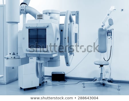 [/rtl] [/rtl]
The heart of an X-ray machine is an electrode pair -- a cathode and an anode -- that sits inside a glass vacuum tube. The cathode is a heated filament, like you might find in an older fluorescent lamp. The machine passes current through the filament, heating it up. The heat sputters electrons off of the filament surface. The positively-charged anode, a flat disc made of tungsten, draws the electrons across the tube.
The voltage difference between the cathode and anode is extremely high, so the electrons fly through the tube with a great deal of force. When a speeding electron collides with a tungsten atom, it knocks loose an electron in one of the atom's lower orbitals. An electron in a higher orbital immediately falls to the lower energy level, releasing its extra energy in the form of a photon. It's a big drop, so the photon has a high energy level -- it is an X-ray photon.
The free electron collides with the tungsten atom, knocking an electron out of a lower orbital. A higher orbital electron fills the empty position, releasing its excess energy as a photon.
Free electrons can also generate photons without hitting an atom. An atom's nucleus may attract a speeding electron just enough to alter its course. Like a comet whipping around the sun, the electron slows down and changes direction as it speeds past the atom. This "braking" action causes the electron to emit excess energy in the form of an X-ray photon.
The free electron is attracted to the tungsten atom nucleus. As the electron speeds past, the nucleus alters its course. The electron loses energy, which it releases as an X-ray photon.
Contrast Media
In a normal X-ray picture, most soft tissue doesn't show up clearly. To focus in on organs, or to examine the blood vessels that make up the circulatory system, doctors must introduce contrast media into the body.
Contrast media are liquids that absorb X-rays more effectively than the surrounding tissue. To bring organs in the digestive and endocrine systems into focus, a patient will swallow a contrast media mixture, typically a barium compound.
If the doctors want to examine blood vessels or other elements in the circulatory system, they will inject contrast media into the patient's bloodstream.
Contrast media are often used in conjunction with a fluoroscope. In fluoroscopy, the X-rays pass through the **** onto a fluorescent screen, creating a moving X-ray image.
Doctors may use fluoroscopy to trace the passage of contrast media through the ****.
Doctors can also record the moving X-ray images on film or video.
The high-impact collisions involved in X-ray production generate a lot of heat. A motor rotates the anode to keep it from melting (the electron beam isn't always focused on the same area). A cool oil bath surrounding the envelope also absorbs heat.
The entire mechanism is surrounded by a thick lead shield. This keeps the X-rays from escaping in all directions. A small ****** in the shield lets some of the X-ray photons escape in a narrow beam. The beam passes through a series of filters on its way to the patient.
A camera on the other side of the patient records the pattern of X-ray light that passes all the way through the patient's ****. The X-ray camera uses the same film technology as an ordinary camera, but X-ray light sets off the chemical reaction instead of visible light.
Generally, doctors keep the film image as a negative. That is, the areas that are exposed to more light appear darker and the areas that are exposed to less light appear lighter. Hard material, such as bone, appears white, and softer material appears black or gray. Doctors can bring different materials into focus by varying the intensity of the X-ray beam.
__________________ | |
| | | | | | كل شئ عن الأشعه السينيه | |
|
| | صلاحيات هذا المنتدى: | لاتستطيع الرد على المواضيع في هذا المنتدى
| |
| |
| |
|


 [/rtl]
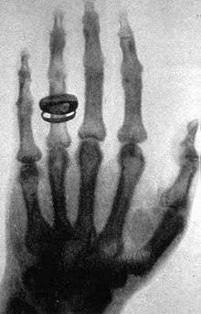 [/rtl]
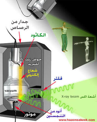 [/rtl]
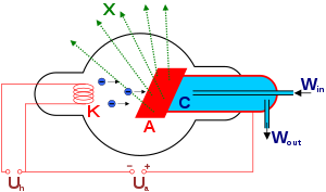 [/rtl]
 [/rtl]